Saprobic on Lonicera maackii. Morph seksual tidak ditentukan. Morph asexual konidiomata 95–150 μm tinggi, 110–170 μm berlian, pycnidial, semi-meremet, terlihat seperti naik, titik hitam di permukaan inang, solitary, globose ke subglobosa, glabrous, uni-loculate, ostiolate di tengah, dengan papilla kecil, tidak memiliki perifise. Conidiomata walls 5–15 μm diamond, dinding tipis, tebalnya tidak sama, sedikit tebal di dasar, terdiri dari lapisan 5–7, sel-sel pseudoparenkimat berwarna coklat yang rata, sedikit gelap di puncak, diatur dalam tekstura angularis hingga tekstura prismatica.
Profil ini diimpor dari uraian spesies pada sumber berlisensi dan sebagian teks diterjemahkan otomatis. Pemeriksaan editorial per profil belum selesai. Bagian yang belum diterjemahkan atau belum lolos pemeriksaan terjemahan tetap ditampilkan dalam bahasa sumber. Angka dan nama ilmiah dipertahankan.
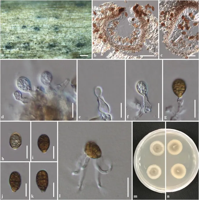
Saprobic on Lonicera maackii. Sexual morph Undetermined. Asexual morph Conidiomata 95–150 μm high, 110–170 μm diam., pycnidial, semi-immersed, visible as raised, black dots on host surface, solitary, globose to subglobose, glabrous, uni-loculate, ostiolate at centre, with minute papilla, lacking periphyses. Conidiomata walls 5–15 μm diam., thin-walled, of unequal thickness, slightly thickened at the base, composed of 5–7 layers, of flattened, brown pseudoparenchymatous cells, slightly dark at the apex, arranged in textura angularis to textura prismatica. Conidiophores reduced to conidiogenous cells. Conidiogenous cells 8–15 × (3–)4–8 µm (\(\bar{x} \) = 11 × 5.5 μm, n = 35), enteroblastic, phialidic, rarely annellidic, discrete, determinate, hyaline, smooth, aseptate, cylindrical to doliiform, with narrow channel, minute collarette and periclinal wall thickening, arising from the inner cavity of pycnidial wall. Conidia (13.5–)14–17(–18.5) × 7–10(–12) µm (\(\bar{x} \) = 15.6 × 9.4 μm, n = 50), initially light brown to pale yellowish, aseptate, becoming reddish brown to dark brown, muriform, subglobose to obovoid, or turbinate, with truncate base, (1–)2–4 transverse septa, with several longitudinal sectors, not constricted at the septa, smooth-walled with minute guttules. Culture characteristics: Colonies on PDA reaching 30–35 mm diam. after 3 weeks at 20–25 °C; colony from above, white-grey at the margin, grey at the centre; from below, white-grey at the margin, grey to dark grey at the centre, slightly radiated outwards colony; dense, circular, slightly raised to umbonate, surface smooth, with edge entire, floccose; not producing pigmentation in agar.
Sebagian uraian ditampilkan dalam bahasa sumber; terjemahannya belum tersedia atau belum lolos pemeriksaan.
Pengukuran mengikuti objek, spesimen atau kultur, dan metode yang disebutkan sumber. Baca uraian asli untuk konteks lengkap.
02 / Kehidupan
Habitat dan peran ekologis
Saprobic on Lonicera maackii. Sexual morph Undetermined. Asexual morph Conidiomata 95–150 μm high, 110–170 μm diam., pycnidial, semi-immersed, visible as raised, black dots on host surface, solitary, globose to subglobose, glabrous, uni-loculate, ostiolate at centre, with minute papilla, lacking periphyses. Conidiomata walls 5–15 μm diam., thin-walled, of unequal thickness, slightly thickened at the base, composed of 5–7 layers, of flattened, brown pseudoparenchymatous cells, slightly dark at the apex, arranged in textura angularis to textura prismatica. Conidiophores reduced to conidiogenous cells. Conidiogenous cells 8–15 × (3–)4–8 µm (\(\bar{x} \) = 11 × 5.5 μm, n = 35), enteroblastic, phialidic, rarely annellidic, discrete, determinate, hyaline, smooth, aseptate, cylindrical to doliiform, with narrow channel, minute collarette and periclinal wall thickening, arising from the inner cavity of pycnidial wall. Conidia (13.5–)14–17(–18.5) × 7–10(–12) µm (\(\bar{x} \) = 15.6 × 9.4 μm, n = 50), initially light brown to pale yellowish, aseptate, becoming reddish brown to dark brown, muriform, subglobose to obovoid, or turbinate, with truncate base, (1–)2–4 transverse septa, with several longitudinal sectors, not constricted at the septa, smooth-walled with minute guttules.
Sebagian uraian ditampilkan dalam bahasa sumber; terjemahannya belum tersedia atau belum lolos pemeriksaan.
Saprobic on Lonicera maackii. Sexual morph Undetermined. Asexual morph Conidiomata 95–150 μm high, 110–170 μm diam., pycnidial, semi-immersed, visible as raised, black dots on host surface, solitary, globose to subglobose, glabrous, uni-loculate, ostiolate at centre, with minute papilla, lacking periphyses. Conidiomata walls 5–15 μm diam., thin-walled, of unequal thickness, slightly thickened at the base, composed of 5–7 layers, of flattened, brown pseudoparenchymatous cells, slightly dark at the apex, arranged in textura angularis to textura prismatica. Conidiophores reduced to conidiogenous cells. Conidiogenous cells 8–15 × (3–)4–8 µm (\(\bar{x} \) = 11 × 5.5 μm, n = 35), enteroblastic, phialidic, rarely annellidic, discrete, determinate, hyaline, smooth, aseptate, cylindrical to doliiform, with narrow channel, minute collarette and periclinal wall thickening, arising from the inner cavity of pycnidial wall.
Sebagian uraian ditampilkan dalam bahasa sumber; terjemahannya belum tersedia atau belum lolos pemeriksaan.
Nama diterima mengikuti Catalogue of Life Base2026-09-11; uraian mempertahankan konsep dan nama sumber aslinya.
Preserve original monograph treatment localities, hosts and specimen/substrate scope. Materials examined document specimens, not the full global range.
Impor dan terjemahan otomatis belum diperiksa editorial per profil. Struktur reproduktif yang disebutkan tidak membuktikan seluruh siklus hidup diketahui.
Pengukuran pada uraian mengikuti spesimen, kultur, dan metode sumber; angka tidak diperluas ke semua populasi.
Catatan distribusi dalam narasi mengikuti sumbernya. Dokumentasi spesimen atau foto mengikuti catatan sumber. Bukti wilayah Indonesia dan penilaian konservasi tidak disimpulkan dari gambar.
05 / Dokumentasi
Melihat dengan konteks
Murilentithecium lonicerae (KUN-HKAS 102238, holotype). a Appearance of conidiomata on host surface. b Section through conidioma. c Section through conidioma wall. d–g Conidiogenous cells and conidia. h–k Conidia. l Germinating of conidium. m, n Culture on PDA after 1 week (m = from above, n = from below). Scale bars a = 200 µm, b = 50 μm, c, l = 20 μm, d–k = 10 μm
Jenis
Pelat ilmiah
Kredit
Penulis dan sumber artikel: Phookamsak, Rungtiwa; Hyde, Kevin D.; Jeewon, Rajesh; Bhat, D. Jayarama; Jones, E. B. Gareth; Maharachchikumbura, Sajeewa S. N.; Raspé, Olivier; Karunarathna, Samantha C.; Wanasinghe, Dhanushka N.; Hongsanan, Sinang; Doilom, Mingkwan; Tennakoon, Danushka S.; Machado, Alexandre R.; Firmino, André L.; Ghosh, Aniket; Karunarathna, Anuruddha; Mešić, Armin; Dutta, Arun Kumar; Thongbai, Benjarong; Devadatha, Bandarupalli; Norphanphoun, Chada; Senwanna, Chanokned; Wei, Deping; Pem, Dhandevi; Ackah, Frank Kwekucher; Wang, Gen-Nuo; Jiang, Hong-Bo; Madrid, Hugo; Lee, Hyang Burm; Goonasekara, Ishani D.; Manawasinghe, Ishara S.; Kušan, Ivana; Cano, Josep; Gené, Josepa; Li, Junfu; Das, Kanad; Acharya, Krishnendu; Raj, K. N. Anil; Latha, K. P. Deepna; Chethana, K. W. Thilini; He, Mao-Qiang; Dueñas, Margarita; Jadan, Margita; Martín, María P.; Samarakoon, Milan C.; Dayarathne, Monika C.; Raza, Mubashar; Park, Myung Soo; Telleria, M. Teresa; Chaiwan, Napalai; Matočec, Neven; de Silva, Nimali I.; Pereira, Olinto L.; Singh, Paras Nath; Manimohan, Patinjareveettil; Uniyal, Priyanka; Shang, Qiu-Ju; Bhatt, Rajendra P.; Perera, Rekhani H.; Alvarenga, Renato Lúcio Mendes; Nogal-Prata, Sandra; Singh, Sanjay K.; Vadthanarat, Santhiti; Oh, Seung-Yoon; Huang, Shi-Ke; Rana, Shiwali; Konta, Sirinapa; Paloi, Soumitra; Jayasiri, Subashini C.; Jeon, Sun Jeong; Mehmood, Tahir; Gibertoni, Tatiana Baptista; Nguyen, Thuong T. T.; Singh, Upendra; Thiyagaraja, Vinodhini; Sarma, V. Venkateswara; Dong, Wei; Yu, Xian-Dong; Lu, Yong-Zhong; Lim, Young Woon; Chen, Yun; Tkalčec, Zdenko; Zhang, Zhi-Feng; Luo, Zong-Long; Daranagama, Dinushani A.; Thambugala, Kasun M.; Tibpromma, Saowaluck; Camporesi, Erio; Bulgakov, Timur S.; Dissanayake, Asha J.; Senanayake, Indunil C.; Dai, Dong Qin; Tang, Li-Zhou; Khan, Sehroon; Zhang, Huang; Promputtha, Itthayakorn; Cai, Lei; Chomnunti, Putarak; Zhao, Rui-Lin; Lumyong, Saisamorn; Boonmee, Saranyaphat; Wen, Ting-Chi; Mortimer, Peter E.; Xu, Jianchu
Saprobic on Lonicera maackii.
Sexual morph Undetermined.
Asexual morph Conidiomata 95–150 μm high, 110–170 μm diam., pycnidial, semi-immersed, visible as raised, black dots on host surface, solitary, globose to subglobose, glabrous, uni-loculate, ostiolate at centre, with minute papilla, lacking periphyses.
Conidiomata walls 5–15 μm diam., thin-walled, of unequal thickness, slightly thickened at the base, composed of 5–7 layers, of flattened, brown pseudoparenchymatous cells, slightly dark at the apex, arranged in textura angularis to textura prismatica.
Saprobic on Lonicera maackii.
Sexual morph Undetermined.
Asexual morph Conidiomata 95–150 μm high, 110–170 μm diam., pycnidial, semi-immersed, visible as raised, black dots on host surface, solitary, globose to subglobose, glabrous, uni-loculate, ostiolate at centre, with minute papilla, lacking periphyses.
Conidiomata walls 5–15 μm diam., thin-walled, of unequal thickness, slightly thickened at the base, composed of 5–7 layers, of flattened, brown pseudoparenchymatous cells, slightly dark at the apex, arranged in textura angularis to textura prismatica.
Conidiophores reduced to conidiogenous cells.
Conidiogenous cells 8–15 × (3–)4–8 µm (\(\bar{x} \) = 11 × 5.5 μm, n = 35), enteroblastic, phialidic, rarely annellidic, discrete, determinate, hyaline, smooth, aseptate, cylindrical to doliiform, with narrow channel, minute collarette and periclinal wall thickening, arising from the inner cavity of pycnidial wall.
Conidia (13.5–)14–17(–18.5) × 7–10(–12) µm (\(\bar{x} \) = 15.6 × 9.4 μm, n = 50), initially light brown to pale yellowish, aseptate, becoming reddish brown to dark brown, muriform, subglobose to obovoid, or turbinate, with truncate base, (1–)2–4 transverse septa, with several longitudinal sectors, not constricted at the septa, smooth-walled with minute guttules.
Culture characteristics: Colonies on PDA reaching 30–35 mm diam. after 3 weeks at 20–25 °C; colony from above, white-grey at the margin, grey at the centre; from below, white-grey at the margin, grey to dark grey at the centre, slightly radiated outwards colony; dense, circular, slightly raised to umbonate, surface smooth, with edge entire, floccose; not producing pigmentation in agar.
Saprobic on Lonicera maackii.
Sexual morph Undetermined.
Asexual morph Conidiomata 95–150 μm high, 110–170 μm diam., pycnidial, semi-immersed, visible as raised, black dots on host surface, solitary, globose to subglobose, glabrous, uni-loculate, ostiolate at centre, with minute papilla, lacking periphyses.
Conidiomata walls 5–15 μm diam., thin-walled, of unequal thickness, slightly thickened at the base, composed of 5–7 layers, of flattened, brown pseudoparenchymatous cells, slightly dark at the apex, arranged in textura angularis to textura prismatica.
Conidiophores reduced to conidiogenous cells.
Conidiogenous cells 8–15 × (3–)4–8 µm (\(\bar{x} \) = 11 × 5.5 μm, n = 35), enteroblastic, phialidic, rarely annellidic, discrete, determinate, hyaline, smooth, aseptate, cylindrical to doliiform, with narrow channel, minute collarette and periclinal wall thickening, arising from the inner cavity of pycnidial wall.
Conidia (13.5–)14–17(–18.5) × 7–10(–12) µm (\(\bar{x} \) = 15.6 × 9.4 μm, n = 50), initially light brown to pale yellowish, aseptate, becoming reddish brown to dark brown, muriform, subglobose to obovoid, or turbinate, with truncate base, (1–)2–4 transverse septa, with several longitudinal sectors, not constricted at the septa, smooth-walled with minute guttules.
Saprobic on Lonicera maackii.
Sexual morph Undetermined.
Asexual morph Conidiomata 95–150 μm high, 110–170 μm diam., pycnidial, semi-immersed, visible as raised, black dots on host surface, solitary, globose to subglobose, glabrous, uni-loculate, ostiolate at centre, with minute papilla, lacking periphyses.
Conidiomata walls 5–15 μm diam., thin-walled, of unequal thickness, slightly thickened at the base, composed of 5–7 layers, of flattened, brown pseudoparenchymatous cells, slightly dark at the apex, arranged in textura angularis to textura prismatica.
Conidiophores reduced to conidiogenous cells.
Conidiogenous cells 8–15 × (3–)4–8 µm (\(\bar{x} \) = 11 × 5.5 μm, n = 35), enteroblastic, phialidic, rarely annellidic, discrete, determinate, hyaline, smooth, aseptate, cylindrical to doliiform, with narrow channel, minute collarette and periclinal wall thickening, arising from the inner cavity of pycnidial wall.
Status: impor sumber; pemeriksaan editorial per profil belum selesai. Untuk klaim biologis, gunakan rujukan pada bagian terkait dan periksa konteks sumber aslinya.
Culture characteristics: Ascospore germinating pada PDA dalam 12 h. Koloni pada PDA mencapai diameter 3 cm dalam minggu 6 pada suhu kamar. Mycelium superficial, awalnya putih, kemudian menjadi cokelat ke hitam, dengan mycelium udara padat…
Saprobic on batang mati dari Centaurea L. Morph seksual Ascomata 170–240 × 270–320 μm (\(\bar{x} \) = 165 × 240 μm, n = 5), di permukaan inang, ditutupi oleh pseudoclypeus, terendam dalam permukaan, sepi, tersebar, uni-lokulat, obpyriform…
Saprobic on tulang rusuk udara kering dari Rosa canina. Morph seksual tidak ditentukan. Morph asexual Conidiomata up ke 140 μm diam., terendam, pycnidial, bercahaya, cokelat ke cokelat gelap, globlosus ke subglobosa, uni-loculate,…