Ciri dan struktur
Saprobic on decaying branches of Areca sp. Sexual morph: Undetermined. Asexual morph: Hyphomycetous (in vivo, Fig. 7). Colonies on natural substrates hairy, black, with glistering white conidial masses. Conidiophores 108–503 × 3–7 µm (x– = 252 × 4.5 μm, n = 15), solitary or in groups, macronematous, mononematous, mostly straight or slightly flexuous or bent, wider at the base, unbranched or branched, septate, thick-walled, smooth-walled or slightly asperulate, brown, pale brown to subhyaline or hyaline near the apex, bearing whorls of 3–6(–7) phialides. Conidiogenous cells 13.5–27 × 2–3 µm (x– = 19 × 2.5 μm, n = 20), monophialidic, subhyaline to hyaline, cylindrical, wider at the base and tapering towards the tips, sometimes with an inconspicuous collarette, asperulate. Conidia 3.1−4.8 × 1.4−2.1 µm (x– = 3.9 × 1.8 μm, n = 40), forming in slimy heads, hyaline, cylindrical, obovoid or ellipsoidal, thick-walled, guttulate, verrucose to granular. Hyphomycetous (in vitro, Fig. 8). Colonies on PDA. Vegetative hyphae subhyaline, pale brown to brown, sometimes aggregating into microsclerotium-like structures, moniliform hyphae are present. Conidiophores 178–375 × 2.5–4 µm (x– = 255 × 3 μm, n = 15), arising from vegetative hyphae, mostly straight or slightly flexuous or bent, slightly tapering toward the upper part, unbranched or branched, thick-walled, septate, smooth-walled, sometimes slightly asperulate, brown to pale brown, subhyaline or hyaline at upper part, bearing phialides. Conidiogenous cells 21–35(–43) × 2–4 µm (x– = 29 × 2 μm, n = 20), phialidic, in whorls of 1–6 phialides, smooth-walled, sometimes slightly asperulate, subhyaline to hyaline, cylindrical, wider at the base and tapering towards the tips.
Habitat dan peran ekologis
Saprobic on decaying branches of Areca sp. Sexual morph: Undetermined. Asexual morph: Hyphomycetous (in vivo, Fig. 7). Colonies on natural substrates hairy, black, with glistering white conidial masses. Conidiophores 108–503 × 3–7 µm (x– = 252 × 4.5 μm, n = 15), solitary or in groups, macronematous, mononematous, mostly straight or slightly flexuous or bent, wider at the base, unbranched or branched, septate, thick-walled, smooth-walled or slightly asperulate, brown, pale brown to subhyaline or hyaline near the apex, bearing whorls of 3–6(–7) phialides. Conidiogenous cells 13.5–27 × 2–3 µm (x– = 19 × 2.5 μm, n = 20), monophialidic, subhyaline to hyaline, cylindrical, wider at the base and tapering towards the tips, sometimes with an inconspicuous collarette, asperulate. Conidia 3.1−4.8 × 1.4−2.1 µm (x– = 3.9 × 1.8 μm, n = 40), forming in slimy heads, hyaline, cylindrical, obovoid or ellipsoidal, thick-walled, guttulate, verrucose to granular. Hyphomycetous (in vitro, Fig. 8). Colonies on PDA. Vegetative hyphae subhyaline, pale brown to brown, sometimes aggregating into microsclerotium-like structures, moniliform hyphae are present. Conidiophores 178–375 × 2.5–4 µm (x– = 255 × 3 μm, n = 15), arising from vegetative hyphae, mostly straight or slightly flexuous or bent, slightly tapering toward the upper part, unbranched or branched, thick-walled, septate, smooth-walled, sometimes slightly asperulate, brown to pale brown, subhyaline or hyaline at upper part, bearing phialides.
- Habitat atau konteks temuan
- kultur yang didokumentasikan
- Substrat
- kayu
- Peran atau konteks
- Pengurai
Reproduksi
Saprobic on decaying branches of Areca sp. Sexual morph: Undetermined. Asexual morph: Hyphomycetous (in vivo, Fig. 7). Colonies on natural substrates hairy, black, with glistering white conidial masses. Conidiophores 108–503 × 3–7 µm (x– = 252 × 4.5 μm, n = 15), solitary or in groups, macronematous, mononematous, mostly straight or slightly flexuous or bent, wider at the base, unbranched or branched, septate, thick-walled, smooth-walled or slightly asperulate, brown, pale brown to subhyaline or hyaline near the apex, bearing whorls of 3–6(–7) phialides. Conidiogenous cells 13.5–27 × 2–3 µm (x– = 19 × 2.5 μm, n = 20), monophialidic, subhyaline to hyaline, cylindrical, wider at the base and tapering towards the tips, sometimes with an inconspicuous collarette, asperulate.
Catatan identitas dan cakupan
- Nama diterima mengikuti Catalogue of Life Base2026-09-11; uraian mempertahankan konsep dan nama sumber aslinya.
- Preserve original treatment localities, hosts and substrate scope. Materials examined describe documented specimens, not the full global range.
- Impor dan terjemahan otomatis belum diperiksa editorial per profil. Struktur reproduktif yang disebutkan tidak membuktikan seluruh siklus hidup diketahui.
- Pengukuran pada uraian mengikuti spesimen, kultur, dan metode sumber; angka tidak diperluas ke semua populasi.
Catatan distribusi dalam narasi mengikuti sumbernya. Dokumentasi spesimen atau foto mengikuti catatan sumber. Bukti wilayah Indonesia dan penilaian konservasi tidak disimpulkan dari gambar.
Melihat dengan konteks
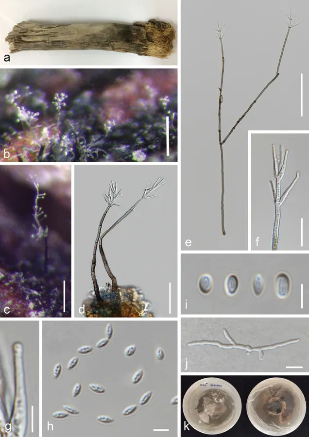
Musicillium theobromae (MFLU 24-0454, a new host record) a appearance of colony on the host b, c a close appearance of colony on the host d, e conidiophores f conidiogenous cells and developing conidia g close up of the phialide with an inconspicuous collarette h, i conidia j germinated conidium k colony on PDA after two months. Scale bars: 100 µm (b,e); 200 µm (c); 50 µm (d); 20 µm (f); 5 µm (g–i); 10 µm (j).
- Jenis
- Pelat ilmiah
- Kredit
- Raheleh Asghari, Chayanard Phukhamsakda, E. B. Gareth Jones, Ali Bahkali, Carlo Chris S. Apurillo, Omid Karimi, Pattana Kakumyan, Kevin D. Hyde
- Lisensi
- CC BY 4.0
- Sumber media
- Buka entri sumber
Dokumentasi tambahan
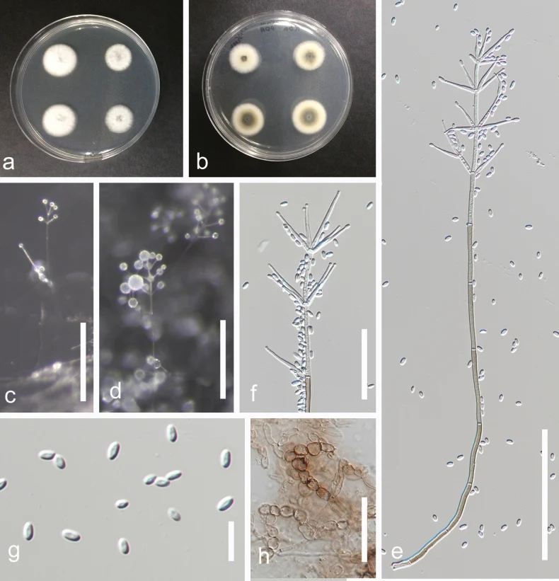
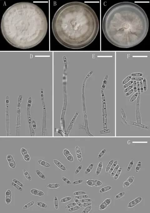
Musicillium theobromae (MFLUCC 24-0563, a new host record) a colony on PDA after two weeks (above) b colony on PDA after two weeks (below) c, d appearance of conidiophores and conidia on the culture e conidiophore and conidia f conidiogenous cells bearing conidia g conidia h moniliform hyphae. Scale bars: 200 µm (c); 100 µm (d, e); 50 µm (f,h); 10 µm (g).
Kredit dan penggunaan
Raheleh Asghari, Chayanard Phukhamsakda, E. B. Gareth Jones, Ali Bahkali, Carlo Chris S. Apurillo, Omid Karimi, Pattana Kakumyan, Kevin D. Hyde
Ukuran dan format gambar disesuaikan tanpa pemotongan, retus, atau penambahan skala. Lisensi sumber dipertahankan.
Sumber profil
- [1] Morphology and phylogeny reveal two new species and host records of hyphomycetous fungi on Areca species from marine habitats in Thailand — Musicillium theobromae
- [2] Catalogue of Life Base2026-09-11 — Musicillium theobromae
- [3] Dokumentasi visual — Musicillium theobromae
- [4] Dokumentasi visual — Musicillium theobromae
Rujukan halaman ini
Fungi. Musicillium theobromae. Rilis data fungi-2026-10-12-expansion-2. https://fungi-atlas.mgadib.chatgpt.site/jamur/musicillium-theobromae/.
Status: impor sumber; pemeriksaan editorial per profil belum selesai. Untuk klaim biologis, gunakan rujukan pada bagian terkait dan periksa konteks sumber aslinya.