Ciri dan struktur
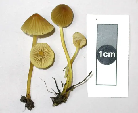
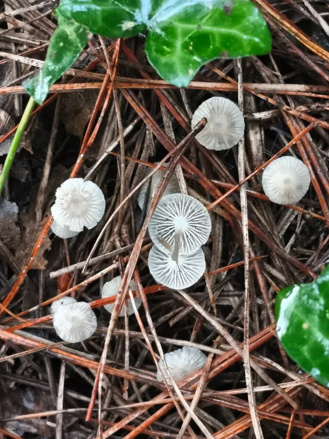
The cap of M. cinerella is white and small, with a diameter typically ranging from 0.5 to 1.5 cm. Initially hemispherical, obtusely conic, and then convex, it expands during maturity, developing visible grooves on the surface that correspond to the gills underneath the cap. The mushroom is inedible. The spores are 7–9 by 4–5 μm, ellipsoid, smooth, and amyloid (reaction very weak in some collections). The basidia (spore-bearing cells) are four-spored, or occasionally a combination of two- and four-spored. The four-spored forms have clamp connections that are absent in the two-spored forms. The pleurocystidia (cystidia found on the face of the gills) are not differentiated. The cheilocystidia (cystidia found on the edges of the gills) are embedded in the hymenium and inconspicuous, measuring 22–36 by 5–11 μm. They are roughly filiform (like thin filaments), with numerous contorted branches or protuberances, and club-shaped with finger-like prolongations. The flesh of the gills is homogeneous, and turns vinaceous-brown when stained in iodine. The flesh of the cap has a well-differentiated pellicle, with a differentiated but not very well-developed hypoderm, and hyphae that are 10–20 μm wide.
Habitat dan peran ekologis
Mycena cinerella adalah jamur saprobik, yang memperoleh nutrisi dengan membusuk sampah daun dan detritus serupa, mengubahnya menjadi humus dan mengmineralisasi bahan organik di tanah. Tubuh buah tumbuh dalam kelompok pada jarum di bawah pinus dan Douglas fir, biasanya pada akhir musim panas dan musim gugur. Di Amerika Serikat, telah dikumpulkan dari Michigan, Washington, Oregon, dan California. Di Eropa, juga telah dikumpulkan dari Inggris, Norwegia, Polandia, dan Swedia.
- Habitat atau konteks temuan
- Informasi belum tersedia pada sumber rilis ini.
- Substrat
- Informasi belum tersedia pada sumber rilis ini.
- Peran atau konteks
- Pengurai
Reproduksi
The spores are 7–9 by 4–5 μm, ellipsoid, smooth, and amyloid (reaction very weak in some collections). The basidia (spore-bearing cells) are four-spored, or occasionally a combination of two- and four-spored. The four-spored forms have clamp connections that are absent in the two-spored forms. The pleurocystidia (cystidia found on the face of the gills) are not differentiated. The cheilocystidia (cystidia found on the edges of the gills) are embedded in the hymenium and inconspicuous, measuring 22–36 by 5–11 μm. They are roughly filiform (like thin filaments), with numerous contorted branches or protuberances, and club-shaped with finger-like prolongations. The flesh of the gills is homogeneous, and turns vinaceous-brown when stained in iodine.
Catatan identitas dan cakupan
- Nama diterima mengikuti Catalogue of Life Base2026-09-11; uraian mempertahankan konsep dan nama sumber aslinya.
- Original geographic, specimen and life-cycle scope retained; source import awaits individual editorial review.
- Impor dan terjemahan otomatis belum diperiksa editorial per profil. Struktur reproduktif yang disebutkan tidak membuktikan seluruh siklus hidup diketahui.
- Pengukuran pada uraian mengikuti spesimen, kultur, dan metode sumber; angka tidak diperluas ke semua populasi.
Catatan distribusi dalam narasi mengikuti sumbernya. Dokumentasi spesimen atau foto mengikuti catatan sumber. Bukti wilayah Indonesia dan penilaian konservasi tidak disimpulkan dari gambar.
Melihat dengan konteks
Dokumentasi Mycena cinerella menurut asosiasi foto pada record takson iNaturalist. Kredit: Jose Castro. Identitas mengikuti sumber; bukan penentuan spesimen independen.
- Jenis
- Fotografi
- Kredit
- (c) Jose Castro, some rights reserved (CC BY)
- Lisensi
- CC BY 4.0
- Sumber media
- Buka entri sumber
Sumber profil
- [1] Mycena cinerella — Wikipedia (en)
- [2] Catalogue of Life Base2026-09-11 — Mycena cinerella
- [3] Dokumentasi visual — Mycena cinerella
Rujukan halaman ini
Fungi. Mycena cinerella. Rilis data fungi-2026-10-12-expansion-2. https://fungi-atlas.mgadib.chatgpt.site/jamur/mycena-cinerella/.
Status: impor sumber; pemeriksaan editorial per profil belum selesai. Untuk klaim biologis, gunakan rujukan pada bagian terkait dan periksa konteks sumber aslinya.