Ciri dan struktur
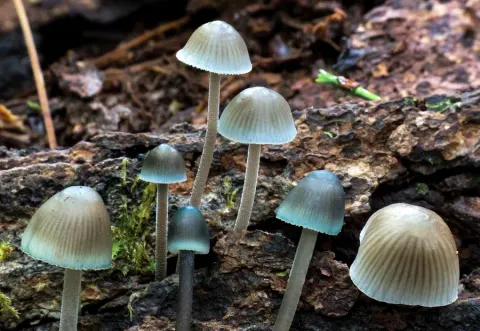
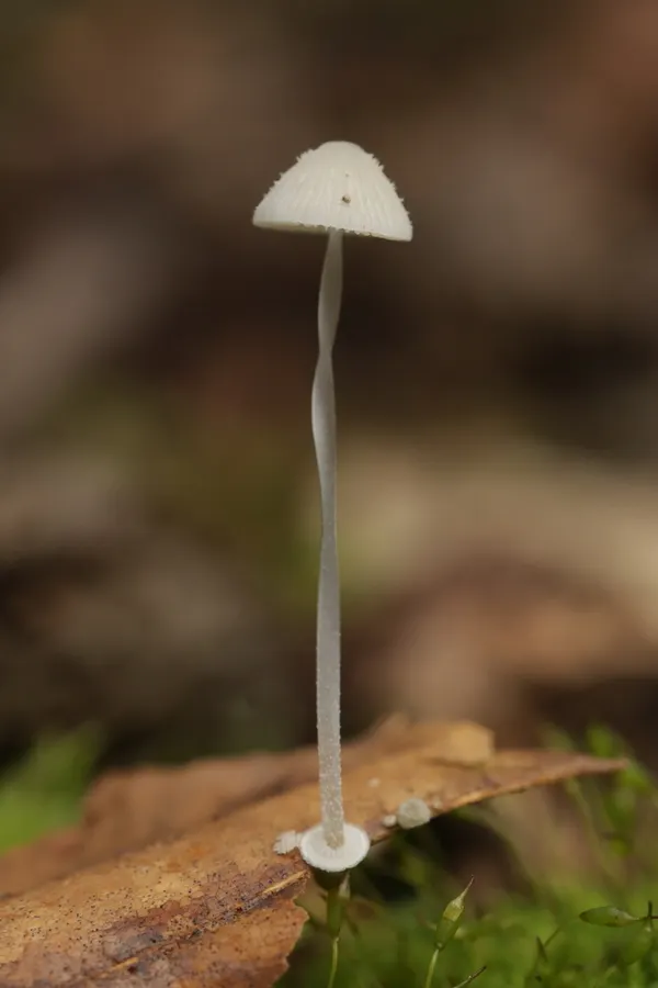
The cap of M. stylobates is 3 – 15 mm in diameter, and depending on its age may range in shape from obtusely conic to convex to bell-shaped to flattened. The structure of the cap margin also depends on the age of the mushroom, progressing from straight or curved inward slightly, to margin flaring or curved backward. The cap surface is smooth, although if viewed with a magnifying glass, minute spines can be seen. As it ages, the surface becomes smooth, moist and somewhat glistening, and it shows grooves corresponding to the position of the gills underneath the cap. The cap color is evenly pale watery gray. The flesh is thin, pallid, and has no distinguishable odor or taste. The insubstantial fruit bodies are considered inedible. The spores are 6–10 by 3.5–4.5 μm, narrowly ellipsoid, and faintly amyloid. The basidia (spore-bearing cells) are four-spored, rarely two-spored. The pleurocystidia (cystidia on the gill face) are not differentiated. The cheilocystidium (cystidia on the gill edge) are abundant and variable in structure, usually club-shaped with between two and five thick obtuse projections that arise from near the apex, sometimes more or less covered with numerous protuberances over the enlarged portion and the neck more or less contorted. They measure 26–38 by 8–13 μm, and are hyaline. The gill flesh is made of greatly enlarged cells, and stains pale vinaceous (red wine color) in iodine. The flesh of the cap has a pellicle which usually gelatinizes in potassium hydroxide or water mounts prepared for microscopy. The surface hyphae are covered with short rodlike projections.
Habitat dan peran ekologis
The fruit bodies of Mycena stylobates grow scattered or in groups on oak leaves or coniferous needles, in the spring and summer or early autumn. Hal ini umum terjadi pada musim panas dan basah. Mycena specialist Alexander H. Smith has mengumpulkan di Tennessee, Michigan, Idaho, dan Washington in Amerika Serikat, dan di Nova Scotia and Ontario in Kanada. Hal ini juga ditemukan di Eropa, termasuk Inggris, Denmark, Jerman, Norwegia, Polandia, Rumania, Skotlandia, Serbia, Swedia. Although it telah dilaporkan beberapa kali dari Australia, ahli mikologi Cheryl Grgurinovic concluded dalam sebuah publikasi 2003 bahwa catatan "sebaiknya dianggap salah".
- Habitat atau konteks temuan
- Informasi belum tersedia pada sumber rilis ini.
- Substrat
- Informasi belum tersedia pada sumber rilis ini.
- Peran atau konteks
- Belum dikategorikan pada rilis ini.
Reproduksi
The spores are 6–10 by 3.5–4.5 μm, narrowly ellipsoid, and faintly amyloid. The basidia (spore-bearing cells) are four-spored, rarely two-spored. The pleurocystidia (cystidia on the gill face) are not differentiated. The cheilocystidium (cystidia on the gill edge) are abundant and variable in structure, usually club-shaped with between two and five thick obtuse projections that arise from near the apex, sometimes more or less covered with numerous protuberances over the enlarged portion and the neck more or less contorted. They measure 26–38 by 8–13 μm, and are hyaline. The gill flesh is made of greatly enlarged cells, and stains pale vinaceous (red wine color) in iodine. The flesh of the cap has a pellicle which usually gelatinizes in potassium hydroxide or water mounts prepared for microscopy.
Catatan identitas dan cakupan
- Nama diterima mengikuti Catalogue of Life Base2026-09-11; uraian mempertahankan konsep dan nama sumber aslinya.
- Original geographic, specimen and life-cycle scope retained; source import awaits individual editorial review.
- Impor dan terjemahan otomatis belum diperiksa editorial per profil. Struktur reproduktif yang disebutkan tidak membuktikan seluruh siklus hidup diketahui.
- Pengukuran pada uraian mengikuti spesimen, kultur, dan metode sumber; angka tidak diperluas ke semua populasi.
Catatan distribusi dalam narasi mengikuti sumbernya. Dokumentasi spesimen atau foto mengikuti catatan sumber. Bukti wilayah Indonesia dan penilaian konservasi tidak disimpulkan dari gambar.
Melihat dengan konteks
Dokumentasi Mycena stylobates menurut asosiasi foto pada record takson iNaturalist. Kredit: Björn Sothmann. Identitas mengikuti sumber; bukan penentuan spesimen independen.
- Jenis
- Fotografi
- Kredit
- (c) Björn Sothmann, some rights reserved (CC BY-SA), uploaded by Björn Sothmann
- Lisensi
- CC BY-SA 4.0
- Sumber media
- Buka entri sumber
Dokumentasi tambahan
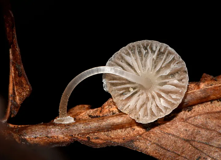
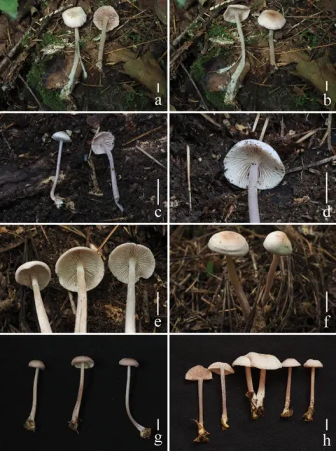
Foto yang diasosiasikan dengan Mycena stylobates pada iNaturalist. Identifikasi mengikuti sumber; bukan penentuan spesimen independen.
Kredit dan penggunaan
(c) Alan Rockefeller, some rights reserved (CC BY), uploaded by Alan Rockefeller
Ukuran dan format gambar disesuaikan tanpa pemotongan, retus, atau penambahan skala. Lisensi sumber dipertahankan.
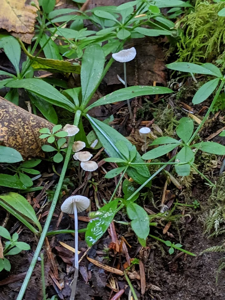
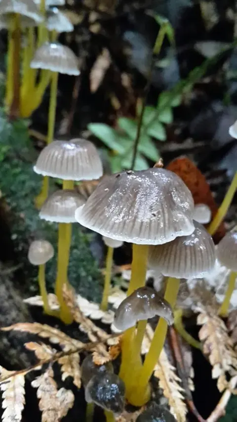
Foto yang diasosiasikan dengan Mycena stylobates pada iNaturalist. Identifikasi mengikuti sumber; bukan penentuan spesimen independen.
Kredit dan penggunaan
(c) Sharon Squazzo, some rights reserved (CC BY), uploaded by Sharon Squazzo
Ukuran dan format gambar disesuaikan tanpa pemotongan, retus, atau penambahan skala. Lisensi sumber dipertahankan.
Sumber profil
- [1] Mycena stylobates — Wikipedia (en)
- [2] Catalogue of Life Base2026-09-11 — Mycena stylobates
- [3] Dokumentasi visual — Mycena stylobates
- [4] Dokumentasi visual — Mycena stylobates
- [5] Dokumentasi visual — Mycena stylobates
Rujukan halaman ini
Fungi. Mycena stylobates. Rilis data fungi-2026-10-12-expansion-2. https://fungi-atlas.mgadib.chatgpt.site/jamur/mycena-stylobates/.
Status: impor sumber; pemeriksaan editorial per profil belum selesai. Untuk klaim biologis, gunakan rujukan pada bagian terkait dan periksa konteks sumber aslinya.