Ciri dan struktur
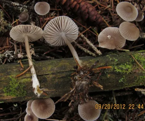
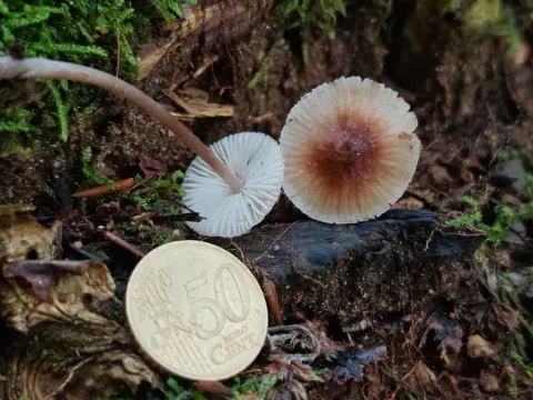
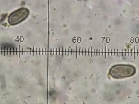
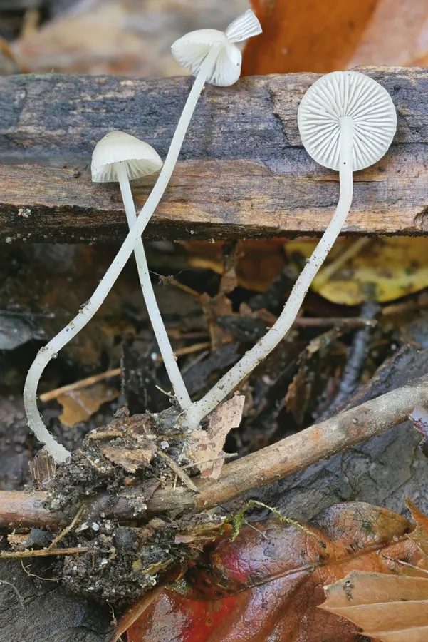
The cap of M. vitilis is initially conic or bell-shaped, but flattens out in maturity, and typically reaches dimensions of up to 2.2 cm. The mushroom is inedible. The spores are ellipsoid, hyaline, amyloid, and measure 9–11 by 5–6 μm. The basidia (spore-bearing cells) are four-spored. The pleurocystidia (cystidia on the gill face) are not differentiated or are occasionally present near the gill edge and similar to cheilocystidia (cystidia on the gill edge). The cheilocystidia, which measure 32–46 by 8–14 μm, are tapered on either end and can have two to several obtuse fingerlike projections arising from the apex. The gill flesh stains vinaceous-brown in iodine. The subhymenium (the tissue layer directly underneath the hymenium) is made of narrow, interwoven hyphae, with the central portion composed of long, cylindrical, and moderately broad cells. The flesh of the cap has a fairly thick subgelatinous pellicle, a well-differentiated hypoderm, and a filamentous tramal body. All except the pellicle stain vinaceous-brown in iodine.
Habitat dan peran ekologis
The fruit bodies of Mycena vitilis are found growing scattered or in groups on debris in hardwood or mixed conifer and hardwood forests. Along the Pasifik Coast it kadang-kadang banyak di Red alder potongan. Di Timur Utara America it cukup umum ditemukan tumbuh di bulan musim gugur October and November with M. semivestipes dan M. pullata. Jamur ini tersebar luas di Eropa (misalnya, Inggris, Jerman, Italia, Norwegia, Polandia, Portugal).
- Habitat atau konteks temuan
- hutan
- Substrat
- Informasi belum tersedia pada sumber rilis ini.
- Peran atau konteks
- Belum dikategorikan pada rilis ini.
Reproduksi
The spores are ellipsoid, hyaline, amyloid, and measure 9–11 by 5–6 μm. The basidia (spore-bearing cells) are four-spored. The pleurocystidia (cystidia on the gill face) are not differentiated or are occasionally present near the gill edge and similar to cheilocystidia (cystidia on the gill edge). The cheilocystidia, which measure 32–46 by 8–14 μm, are tapered on either end and can have two to several obtuse fingerlike projections arising from the apex. The gill flesh stains vinaceous-brown in iodine. The subhymenium (the tissue layer directly underneath the hymenium) is made of narrow, interwoven hyphae, with the central portion composed of long, cylindrical, and moderately broad cells. The flesh of the cap has a fairly thick subgelatinous pellicle, a well-differentiated hypoderm, and a filamentous tramal body.
Catatan identitas dan cakupan
- Nama diterima mengikuti Catalogue of Life Base2026-09-11; uraian mempertahankan konsep dan nama sumber aslinya.
- Original geographic, specimen and life-cycle scope retained; source import awaits individual editorial review.
- Impor dan terjemahan otomatis belum diperiksa editorial per profil. Struktur reproduktif yang disebutkan tidak membuktikan seluruh siklus hidup diketahui.
- Pengukuran pada uraian mengikuti spesimen, kultur, dan metode sumber; angka tidak diperluas ke semua populasi.
Catatan distribusi dalam narasi mengikuti sumbernya. Dokumentasi spesimen atau foto mengikuti catatan sumber. Bukti wilayah Indonesia dan penilaian konservasi tidak disimpulkan dari gambar.
Melihat dengan konteks
Dokumentasi Mycena vitilis menurut asosiasi foto pada record takson iNaturalist. Kredit: Garrett Taylor. Identitas mengikuti sumber; bukan penentuan spesimen independen.
- Jenis
- Fotografi
- Kredit
- no rights reserved, uploaded by Garrett Taylor
- Lisensi
- CC0 1.0
- Sumber media
- Buka entri sumber
Sumber profil
- [1] Mycena vitilis — Wikipedia (en)
- [2] Catalogue of Life Base2026-09-11 — Mycena vitilis
- [3] Dokumentasi visual — Mycena vitilis
Rujukan halaman ini
Fungi. Mycena vitilis. Rilis data fungi-2026-10-12-expansion-2. https://fungi-atlas.mgadib.chatgpt.site/jamur/mycena-vitilis/.
Status: impor sumber; pemeriksaan editorial per profil belum selesai. Untuk klaim biologis, gunakan rujukan pada bagian terkait dan periksa konteks sumber aslinya.