Ciri dan struktur
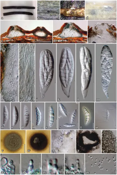
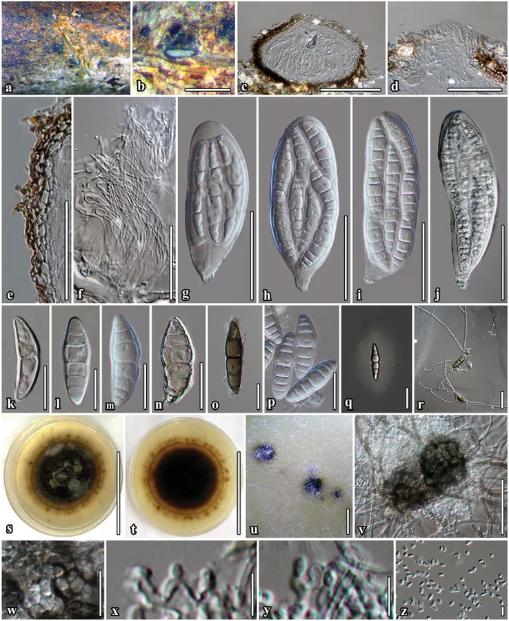
Saprobic on dead twigs of Bauhinia variegata L. Sexual morph. Ascomata 113–190 μm high × 170–307 μm diam. (x̄ = 160 × 260 μm, n = 10), semi-immersed to immersed, solitary, scattered, subglobose to compressed, coriaceous, brown to dark brown, rough-walled, with short hyphae projecting from peridium, ostiolate. Ostiole 33 × 85 μm diam., centrally located, papillate, periphysoid. Peridium 8–25 μm wide (x̄ = 17, n = 30), with cells 3–8 μm wide, composed of 3 layers of reddish-brown to dark brown, cells of textura angularis, inner layer composed of hyaline gelatinous cells. Hamathecium composed of numerous, dense, long, 1–2.4 μm (x̄ = 1.7 μm, n = 50), narrow, filiform, transversely septate, branched, anastomosing, cellular psedoparaphyses. Asci 53–116 × 26–43 μm (x̄ = 98 × 37 μm, n = 30), 8-spored, bitunicate, fissitunicate, oboviod to oblong, with furcate pedicel, with ocular chamber visible when immature. Ascospores 37–46 × 9–16 μm (x̄ = 43 × 13 μm, n = 50), bi-seriate or overlapping, broad fusiform, narrow towards the apex, initially hyaline, becoming brown to dark brown at maturity, 4–7-transversely euseptate, constricted at the septa, with cell above central septum wider, rough-walled, indentations present, surrounded by 7–12 μm wide, mucilaginous sheath. Asexual morph coelomycetous. Pycnidia produced on mycelium in water agar. Conidiomata 33−49 μm high × 92–108 μm wide diam., pycnidial, dark brown to black, covered by dense vegetative hyphae, globose, in agar immersed to superficial, uniloculate, solitary to scattered, ostiolate. Conidiomatal wall thin, brown to black-walled with cells of textura angularis.
Habitat dan peran ekologis
THAILAND. Phrae Province: Song District, on dead twigs of Bauhinia variegata L. (Fabaceae), 25 July 2015, C. Phukhamsakda, S1-11, MFLU 17-0002 (holotype), MFLUCC 16-0398 = ICMP 21572 (ex-type living culture). Saprobic on dead twigs of Bauhinia variegata L. Sexual morph. Ascomata 113–190 μm high × 170–307 μm diam. (x̄ = 160 × 260 μm, n = 10), semi-immersed to immersed, solitary, scattered, subglobose to compressed, coriaceous, brown to dark brown, rough-walled, with short hyphae projecting from peridium, ostiolate. Ostiole 33 × 85 μm diam., centrally located, papillate, periphysoid. Peridium 8–25 μm wide (x̄ = 17, n = 30), with cells 3–8 μm wide, composed of 3 layers of reddish-brown to dark brown, cells of textura angularis, inner layer composed of hyaline gelatinous cells. Hamathecium composed of numerous, dense, long, 1–2.4 μm (x̄ = 1.7 μm, n = 50), narrow, filiform, transversely septate, branched, anastomosing, cellular psedoparaphyses. Asci 53–116 × 26–43 μm (x̄ = 98 × 37 μm, n = 30), 8-spored, bitunicate, fissitunicate, oboviod to oblong, with furcate pedicel, with ocular chamber visible when immature. Ascospores 37–46 × 9–16 μm (x̄ = 43 × 13 μm, n = 50), bi-seriate or overlapping, broad fusiform, narrow towards the apex, initially hyaline, becoming brown to dark brown at maturity, 4–7-transversely euseptate, constricted at the septa, with cell above central septum wider, rough-walled, indentations present, surrounded by 7–12 μm wide, mucilaginous sheath.
- Habitat atau konteks temuan
- kultur yang didokumentasikan
- Substrat
- Informasi belum tersedia pada sumber rilis ini.
- Peran atau konteks
- Pengurai
Reproduksi
Saprobic on dead twigs of Bauhinia variegata L. Sexual morph. Ascomata 113–190 μm high × 170–307 μm diam. (x̄ = 160 × 260 μm, n = 10), semi-immersed to immersed, solitary, scattered, subglobose to compressed, coriaceous, brown to dark brown, rough-walled, with short hyphae projecting from peridium, ostiolate. Ostiole 33 × 85 μm diam., centrally located, papillate, periphysoid. Peridium 8–25 μm wide (x̄ = 17, n = 30), with cells 3–8 μm wide, composed of 3 layers of reddish-brown to dark brown, cells of textura angularis, inner layer composed of hyaline gelatinous cells. Hamathecium composed of numerous, dense, long, 1–2.4 μm (x̄ = 1.7 μm, n = 50), narrow, filiform, transversely septate, branched, anastomosing, cellular psedoparaphyses.
Catatan identitas dan cakupan
- Nama diterima mengikuti Catalogue of Life Base2026-09-11; uraian mempertahankan konsep dan nama sumber aslinya.
- Preserve original treatment localities, hosts and substrate scope. Materials examined describe documented specimens, not the full global range.
- Impor dan terjemahan otomatis belum diperiksa editorial per profil. Struktur reproduktif yang disebutkan tidak membuktikan seluruh siklus hidup diketahui.
- Pengukuran pada uraian mengikuti spesimen, kultur, dan metode sumber; angka tidak diperluas ke semua populasi.
Catatan distribusi dalam narasi mengikuti sumbernya. Dokumentasi spesimen atau foto mengikuti catatan sumber. Bukti wilayah Indonesia dan penilaian konservasi tidak disimpulkan dari gambar.
Melihat dengan konteks
Neoaquastroma bauhiniae (MFLU 17-0002, holotype) a Appearance of ascomata on host surface b Close up of ascoma c Section of ascoma d Ostiolar canal e Section of partial peridium layer f Pseudoparaphyses g–j Development state of asci j Asci produced in culture k–p Development state of ascospores; (n, o Senescent spores m, p ascospores in 5% of KOH reagent); q Ascospores stained with India ink, sheath surrounding the entire ascospore r Germinated ascospore s, t Culture character on MEA u Conidiomata forming on agar on rice straw media after 8 weeks v Immature conidiomata w Conidiomatal wall x, y Conidiogenous cells and developing conidia z Conidia j, m Asci and ascospore in culture (on rice straw). Scale bars: 500 µm (b); 100 µm (c, v); 50 µm (d–j); 20 µm (k–r, w); 5 µm (x–z).
- Jenis
- Pelat ilmiah
- Kredit
- Chayanard Phukhamsakda, Darbhe J. Bhat, Sinang Hongsanan, Jian-Chu Xu, Marc Stadler, Kevin D. Hyde
- Lisensi
- CC BY 4.0
- Sumber media
- Buka entri sumber
Sumber profil
- [1] Two novel species of Neoaquastroma (Parabambusicolaceae, Pleosporales) with their phoma-like asexual morphs — Neoaquastroma bauhiniae
- [2] Catalogue of Life Base2026-09-11 — Neoaquastroma bauhiniae
- [3] Dokumentasi visual — Neoaquastroma bauhiniae
Rujukan halaman ini
Fungi. Neoaquastroma bauhiniae. Rilis data fungi-2026-10-12-expansion-2. https://fungi-atlas.mgadib.chatgpt.site/jamur/neoaquastroma-bauhiniae/.
Status: impor sumber; pemeriksaan editorial per profil belum selesai. Untuk klaim biologis, gunakan rujukan pada bagian terkait dan periksa konteks sumber aslinya.