Ciri dan struktur
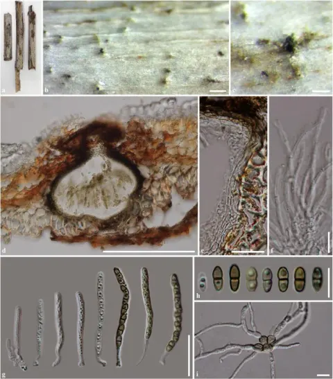
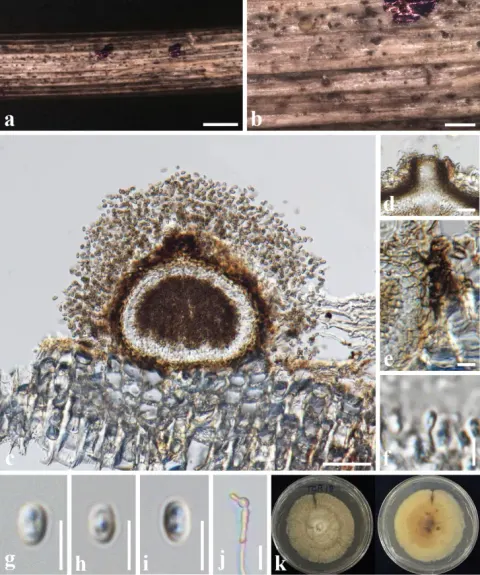
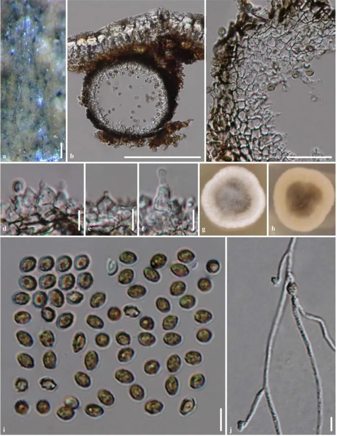
Saprobic on Para rubber (Hevea brasiliensis). Sexual morph Undetermined. Asexual morph Coelomycetous. Conidiomata 90–130 μm high, 115–180 μm diam., pycnidial, immersed, visible as small, brown to black dots on the host surface, solitary, scattered, globose, uni-loculate, glabrous, ostiole not observed. Conidiomata walls 7–15 µm wide, thick-walled, composed of several cell layers of dark brown to black, pseudoparenchymatous cells of textura angularis. Conidiophores reduced to conidiogenous cells. Conidiogenous cells 3–7 × 2–5 μm (\(\bar{x} \) = 5.2 × 3.4 μm, n = 15), lining the inner cavity, difficult to distinguish from pycnidial wall, enteroblastic, phialidic, integrated, simple, short, hyaline, ampulliform to doliiform, thin-walled, smooth. Conidia 2.5–5 × 2–4 µm (\(\bar{x} \) = 4 × 3, n = 50), globose or subglobose to ellipsoidal, initially hyaline, becoming brown to dark brown, aseptate, with one or two guttules, rough-walled, verruculose. Culture characteristics: Colonies on MEA reaching 20–25 mm diam. after 2 weeks at 25–30 °C, colony from above, circular, medium dense, edge entire, velvety to woolly, white at the margin, white to yellowish in the middle, geenish grey to grey at the centre; from below, white at the margin, greenish grey at the centre, with concentric rings radiate.
Habitat dan peran ekologis
Saprobic on Para rubber (Hevea brasiliensis). Sexual morph Undetermined. Asexual morph Coelomycetous. Conidiomata 90–130 μm high, 115–180 μm diam., pycnidial, immersed, visible as small, brown to black dots on the host surface, solitary, scattered, globose, uni-loculate, glabrous, ostiole not observed. Conidiomata walls 7–15 µm wide, thick-walled, composed of several cell layers of dark brown to black, pseudoparenchymatous cells of textura angularis. Conidiophores reduced to conidiogenous cells. Conidiogenous cells 3–7 × 2–5 μm (\(\bar{x} \) = 5.2 × 3.4 μm, n = 15), lining the inner cavity, difficult to distinguish from pycnidial wall, enteroblastic, phialidic, integrated, simple, short, hyaline, ampulliform to doliiform, thin-walled, smooth. Conidia 2.5–5 × 2–4 µm (\(\bar{x} \) = 4 × 3, n = 50), globose or subglobose to ellipsoidal, initially hyaline, becoming brown to dark brown, aseptate, with one or two guttules, rough-walled, verruculose.
- Habitat atau konteks temuan
- Informasi belum tersedia pada sumber rilis ini.
- Substrat
- Informasi belum tersedia pada sumber rilis ini.
- Peran atau konteks
- Pengurai
Reproduksi
Saprobic on Para rubber (Hevea brasiliensis). Sexual morph Undetermined. Asexual morph Coelomycetous. Conidiomata 90–130 μm high, 115–180 μm diam., pycnidial, immersed, visible as small, brown to black dots on the host surface, solitary, scattered, globose, uni-loculate, glabrous, ostiole not observed. Conidiomata walls 7–15 µm wide, thick-walled, composed of several cell layers of dark brown to black, pseudoparenchymatous cells of textura angularis. Conidiophores reduced to conidiogenous cells. Conidiogenous cells 3–7 × 2–5 μm (\(\bar{x} \) = 5.2 × 3.4 μm, n = 15), lining the inner cavity, difficult to distinguish from pycnidial wall, enteroblastic, phialidic, integrated, simple, short, hyaline, ampulliform to doliiform, thin-walled, smooth.
Catatan identitas dan cakupan
- Nama diterima mengikuti Catalogue of Life Base2026-09-11; uraian mempertahankan konsep dan nama sumber aslinya.
- Preserve original monograph treatment localities, hosts and specimen/substrate scope. Materials examined document specimens, not the full global range.
- Impor dan terjemahan otomatis belum diperiksa editorial per profil. Struktur reproduktif yang disebutkan tidak membuktikan seluruh siklus hidup diketahui.
- Pengukuran pada uraian mengikuti spesimen, kultur, dan metode sumber; angka tidak diperluas ke semua populasi.
Catatan distribusi dalam narasi mengikuti sumbernya. Dokumentasi spesimen atau foto mengikuti catatan sumber. Bukti wilayah Indonesia dan penilaian konservasi tidak disimpulkan dari gambar.
Melihat dengan konteks
Neoroussoella heveae (MFLU 17-1983, holotype). a Conidiomata immersed in host substrate. b Section through the conidioma. c Conidioma wall. d–f Conidiogenous cells. g, h Colony on MEA (g = from above, h = from below). i Conidia. j Germinated conidium. Scale bars a = 200 µm, b = 100 µm, c = 20 µm, d–j = 5 µm
- Jenis
- Pelat ilmiah
- Kredit
- Penulis dan sumber artikel: Phookamsak, Rungtiwa; Hyde, Kevin D.; Jeewon, Rajesh; Bhat, D. Jayarama; Jones, E. B. Gareth; Maharachchikumbura, Sajeewa S. N.; Raspé, Olivier; Karunarathna, Samantha C.; Wanasinghe, Dhanushka N.; Hongsanan, Sinang; Doilom, Mingkwan; Tennakoon, Danushka S.; Machado, Alexandre R.; Firmino, André L.; Ghosh, Aniket; Karunarathna, Anuruddha; Mešić, Armin; Dutta, Arun Kumar; Thongbai, Benjarong; Devadatha, Bandarupalli; Norphanphoun, Chada; Senwanna, Chanokned; Wei, Deping; Pem, Dhandevi; Ackah, Frank Kwekucher; Wang, Gen-Nuo; Jiang, Hong-Bo; Madrid, Hugo; Lee, Hyang Burm; Goonasekara, Ishani D.; Manawasinghe, Ishara S.; Kušan, Ivana; Cano, Josep; Gené, Josepa; Li, Junfu; Das, Kanad; Acharya, Krishnendu; Raj, K. N. Anil; Latha, K. P. Deepna; Chethana, K. W. Thilini; He, Mao-Qiang; Dueñas, Margarita; Jadan, Margita; Martín, María P.; Samarakoon, Milan C.; Dayarathne, Monika C.; Raza, Mubashar; Park, Myung Soo; Telleria, M. Teresa; Chaiwan, Napalai; Matočec, Neven; de Silva, Nimali I.; Pereira, Olinto L.; Singh, Paras Nath; Manimohan, Patinjareveettil; Uniyal, Priyanka; Shang, Qiu-Ju; Bhatt, Rajendra P.; Perera, Rekhani H.; Alvarenga, Renato Lúcio Mendes; Nogal-Prata, Sandra; Singh, Sanjay K.; Vadthanarat, Santhiti; Oh, Seung-Yoon; Huang, Shi-Ke; Rana, Shiwali; Konta, Sirinapa; Paloi, Soumitra; Jayasiri, Subashini C.; Jeon, Sun Jeong; Mehmood, Tahir; Gibertoni, Tatiana Baptista; Nguyen, Thuong T. T.; Singh, Upendra; Thiyagaraja, Vinodhini; Sarma, V. Venkateswara; Dong, Wei; Yu, Xian-Dong; Lu, Yong-Zhong; Lim, Young Woon; Chen, Yun; Tkalčec, Zdenko; Zhang, Zhi-Feng; Luo, Zong-Long; Daranagama, Dinushani A.; Thambugala, Kasun M.; Tibpromma, Saowaluck; Camporesi, Erio; Bulgakov, Timur S.; Dissanayake, Asha J.; Senanayake, Indunil C.; Dai, Dong Qin; Tang, Li-Zhou; Khan, Sehroon; Zhang, Huang; Promputtha, Itthayakorn; Cai, Lei; Chomnunti, Putarak; Zhao, Rui-Lin; Lumyong, Saisamorn; Boonmee, Saranyaphat; Wen, Ting-Chi; Mortimer, Peter E.; Xu, Jianchu
- Lisensi
- CC BY 4.0
- Sumber media
- Buka entri sumber
Sumber profil
- [1] Fungal diversity notes 929–1035: taxonomic and phylogenetic contributions on genera and species of fungi — Neoroussoella heveae
- [2] Catalogue of Life Base2026-09-11 — Neoroussoella heveae
- [3] Dokumentasi visual — Neoroussoella heveae
Rujukan halaman ini
Fungi. Neoroussoella heveae. Rilis data fungi-2026-10-12-expansion-2. https://fungi-atlas.mgadib.chatgpt.site/jamur/neoroussoella-heveae/.
Status: impor sumber; pemeriksaan editorial per profil belum selesai. Untuk klaim biologis, gunakan rujukan pada bagian terkait dan periksa konteks sumber aslinya.