Saprobic or lemah patogen pada Prunus mahaleb. Morph seksual tidak ditentukan. Morph asexual konidiomata 220–265 μm tinggi × 260–380 μm diameter. (\(\bar{x} \) = 245 × 340 μm, n = 10), pycnidial, stromatic, solitary atau kluster, semi-mermermerged, erumpent pada kematangan, cokelat gelap ke hitam, globose, tanpa ostiol. Pycnidial walls 25–35 μm lebar, terdiri dari dua lapisan hialin hingga sel-sel pseudoparenkimatus coklat gelap dari tekstura angularis. Conidiogenous cells 10–22 μm tinggi × 4–6 μm lebar, hialin, dinding tipis, holoblastik, halus, silinder, bengkak di dasar, diskrit, menghasilkan satu konidium di puncak.
Profil ini diimpor dari uraian spesies pada sumber berlisensi dan sebagian teks diterjemahkan otomatis. Pemeriksaan editorial per profil belum selesai. Bagian yang belum diterjemahkan atau belum lolos pemeriksaan terjemahan tetap ditampilkan dalam bahasa sumber. Angka dan nama ilmiah dipertahankan.
Saprobic or weak pathogenic on Prunus mahaleb. Sexual morph Undetermined. Asexual morph Conidiomata 220–265 μm high × 260–380 μm diam. (\(\bar{x} \) = 245 × 340 μm, n = 10), pycnidial, stromatic, solitary or clustered, semi-immersed, erumpent at maturity, dark brown to black, globose, without an ostiole. Pycnidial walls 25–35 μm wide, composed of two layers of hyaline to dark brown pseudoparenchymatous cells of textura angularis. Conidiogenous cells 10–22 μm high × 4–6 μm wide, hyaline, thin-walled, holoblastic, smooth, cylindrical, swollen at the base, discrete, producing a single conidium at the apex. Conidia 11–16 × 6–10 μm (\(\bar{x} \) = 14 × 9 μm, n = 50), aseptate, globose to subglobose, widest in the centre, with rounded apex, initially hyaline, becoming dark brown before release from the pycnidia, wall moderately thick, externally smooth, internally roughened. Spermatia 3–5 × 1.5–2 μm, rod-shaped with obtuse ends, hyaline, thin-walled, smooth. Culture characteristics: Conidia germinating on water agar and germ tubes produced from conidia within 12 h from lower end. Colonies growing on PDA, circular, with flat surface, edge entire, reaching 28 mm in 3 weeks at 25 °C, white to pale brown in PDA medium on top. Mycelium superficial and partially immersed, branched, septate, hyaline grey to black, smooth.
Sebagian uraian ditampilkan dalam bahasa sumber; terjemahannya belum tersedia atau belum lolos pemeriksaan.
Pengukuran mengikuti objek, spesimen atau kultur, dan metode yang disebutkan sumber. Baca uraian asli untuk konteks lengkap.
02 / Kehidupan
Habitat dan peran ekologis
Known host and distribution: Saprobic and pathogenic on various hosts from temperate regions (Aveskamp et al. 2010; Chen et al. 2015; Bai et al. 2016; Jiao et al. 2017; Liu et al. 2018b; Moral et al. 2018; Chethana et al. 2019; Farr and Rossman 2020).
Sebagian uraian ditampilkan dalam bahasa sumber; terjemahannya belum tersedia atau belum lolos pemeriksaan.
Saprobic or weak pathogenic on Prunus mahaleb. Sexual morph Undetermined. Asexual morph Conidiomata 220–265 μm high × 260–380 μm diam. (\(\bar{x} \) = 245 × 340 μm, n = 10), pycnidial, stromatic, solitary or clustered, semi-immersed, erumpent at maturity, dark brown to black, globose, without an ostiole. Pycnidial walls 25–35 μm wide, composed of two layers of hyaline to dark brown pseudoparenchymatous cells of textura angularis. Conidiogenous cells 10–22 μm high × 4–6 μm wide, hyaline, thin-walled, holoblastic, smooth, cylindrical, swollen at the base, discrete, producing a single conidium at the apex. Conidia 11–16 × 6–10 μm (\(\bar{x} \) = 14 × 9 μm, n = 50), aseptate, globose to subglobose, widest in the centre, with rounded apex, initially hyaline, becoming dark brown before release from the pycnidia, wall moderately thick, externally smooth, internally roughened.
Sebagian uraian ditampilkan dalam bahasa sumber; terjemahannya belum tersedia atau belum lolos pemeriksaan.
Nama diterima mengikuti Catalogue of Life Base2026-09-11; uraian mempertahankan konsep dan nama sumber aslinya.
Preserve original monograph treatment localities, hosts and specimen/substrate scope. Materials examined document specimens, not the full global range.
Impor dan terjemahan otomatis belum diperiksa editorial per profil. Struktur reproduktif yang disebutkan tidak membuktikan seluruh siklus hidup diketahui.
Pengukuran pada uraian mengikuti spesimen, kultur, dan metode sumber; angka tidak diperluas ke semua populasi.
Catatan distribusi dalam narasi mengikuti sumbernya. Dokumentasi spesimen atau foto mengikuti catatan sumber. Bukti wilayah Indonesia dan penilaian konservasi tidak disimpulkan dari gambar.
05 / Dokumentasi
Melihat dengan konteks
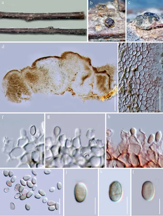
Nothophoma quercina (MFLU 17-2128). a, b Appearance of conidiomata on host surface. c, d Section of conidiomata. e Section through pycnidial wall (stained with congo red). f – h Conidiogenous cells (h = stained with congo red). i – l Conidia stained in congo red. Scale bars: d = 100 µm, e = 30 µm, c, f – l = 10 µm
Jenis
Pelat ilmiah
Kredit
Penulis dan sumber artikel: Hyde, Kevin D.; Dong, Yang; Phookamsak, Rungtiwa; Jeewon, Rajesh; Bhat, D. Jayarama; Jones, E. B. Gareth; Liu, Ning-Guo; Abeywickrama, Pranami D.; Mapook, Ausana; Wei, Deping; Perera, Rekhani H.; Manawasinghe, Ishara S.; Pem, Dhandevi; Bundhun, Digvijayini; Karunarathna, Anuruddha; Ekanayaka, Anusha H.; Bao, Dan-Feng; Li, Junfu; Samarakoon, Milan C.; Chaiwan, Napalai; Lin, Chuan-Gen; Phutthacharoen, Kunthida; Zhang, Sheng-Nan; Senanayake, Indunil C.; Goonasekara, Ishani D.; Thambugala, Kasun M.; Phukhamsakda, Chayanard; Tennakoon, Danushka S.; Jiang, Hong-Bo; Yang, Jing; Zeng, Ming; Huanraluek, Naruemon; Liu, Jian-Kui (Jack); Wijesinghe, Subodini N.; Tian, Qing; Tibpromma, Saowaluck; Brahmanage, Rashika S.; Boonmee, Saranyaphat; Huang, Shi-Ke; Thiyagaraja, Vinodhini; Lu, Yong-Zhong; Jayawardena, Ruvishika S.; Dong, Wei; Yang, Er-Fu; Singh, Sanjay K.; Singh, Shiv Mohan; Rana, Shiwali; Lad, Sneha S.; Anand, Garima; Devadatha, Bandarupalli; Niranjan, M.; Sarma, V. Venkateswara; Liimatainen, Kare; Aguirre-Hudson, Begoña; Niskanen, Tuula; Overall, Andy; Alvarenga, Renato Lúcio Mendes; Gibertoni, Tatiana Baptista; Pfliegler, Walter P.; Horváth, Enikő; Imre, Alexandra; Alves, Amanda Lucia; da Silva Santos, Ana Carla; Tiago, Patricia Vieira; Bulgakov, Timur S.; Wanasinghe, Dhanushaka N.; Bahkali, Ali H.; Doilom, Mingkwan; Elgorban, Abdallah M.; Maharachchikumbura, Sajeewa S. N.; Rajeshkumar, Kunhiraman C.; Haelewaters, Danny; Mortimer, Peter E.; Zhao, Qi; Lumyong, Saisamorn; Xu, Jianchu; Sheng, Jun
Saprobic or weak pathogenic on Prunus mahaleb.
Sexual morph Undetermined.
Asexual morph Conidiomata 220–265 μm high × 260–380 μm diam. (\(\bar{x} \) = 245 × 340 μm, n = 10), pycnidial, stromatic, solitary or clustered, semi-immersed, erumpent at maturity, dark brown to black, globose, without an ostiole.
Pycnidial walls 25–35 μm wide, composed of two layers of hyaline to dark brown pseudoparenchymatous cells of textura angularis.
Conidiogenous cells 10–22 μm high × 4–6 μm wide, hyaline, thin-walled, holoblastic, smooth, cylindrical, swollen at the base, discrete, producing a single conidium at the apex.
Saprobic or weak pathogenic on Prunus mahaleb. Sexual morph Undetermined. Asexual morph Conidiomata 220–265 μm high × 260–380 μm diam. (\(\bar{x} \) = 245 × 340 μm, n = 10), pycnidial, stromatic, solitary or clustered, semi-immersed, erumpent at maturity, dark brown to black, globose, without an ostiole. Pycnidial walls 25–35 μm wide, composed of two layers of hyaline to dark brown pseudoparenchymatous cells of textura angularis. Conidiogenous cells 10–22 μm high × 4–6 μm wide, hyaline, thin-walled, holoblastic, smooth, cylindrical, swollen at the base, discrete, producing a single conidium at the apex. Conidia 11–16 × 6–10 μm (\(\bar{x} \) = 14 × 9 μm, n = 50), aseptate, globose to subglobose, widest in the centre, with rounded apex, initially hyaline, becoming dark brown before release from the pycnidia, wall moderately thick, externally smooth, internally roughened. Spermatia 3–5 × 1.5–2 μm, rod-shaped with obtuse ends, hyaline, thin-walled, smooth.
Culture characteristics: Conidia germinating on water agar and germ tubes produced from conidia within 12 h from lower end. Colonies growing on PDA, circular, with flat surface, edge entire, reaching 28 mm in 3 weeks at 25 °C, white to pale brown in PDA medium on top. Mycelium superficial and partially immersed, branched, septate, hyaline grey to black, smooth.
Known host and distribution: Saprobic and pathogenic on various hosts from temperate regions (Aveskamp et al. 2010; Chen et al. 2015; Bai et al. 2016; Jiao et al. 2017; Liu et al. 2018b; Moral et al. 2018; Chethana et al. 2019; Farr and Rossman 2020).
Saprobic or weak pathogenic on Prunus mahaleb.
Sexual morph Undetermined.
Asexual morph Conidiomata 220–265 μm high × 260–380 μm diam. (\(\bar{x} \) = 245 × 340 μm, n = 10), pycnidial, stromatic, solitary or clustered, semi-immersed, erumpent at maturity, dark brown to black, globose, without an ostiole.
Pycnidial walls 25–35 μm wide, composed of two layers of hyaline to dark brown pseudoparenchymatous cells of textura angularis.
Conidiogenous cells 10–22 μm high × 4–6 μm wide, hyaline, thin-walled, holoblastic, smooth, cylindrical, swollen at the base, discrete, producing a single conidium at the apex.
Conidia 11–16 × 6–10 μm (\(\bar{x} \) = 14 × 9 μm, n = 50), aseptate, globose to subglobose, widest in the centre, with rounded apex, initially hyaline, becoming dark brown before release from the pycnidia, wall moderately thick, externally smooth, internally roughened.
Status: impor sumber; pemeriksaan editorial per profil belum selesai. Untuk klaim biologis, gunakan rujukan pada bagian terkait dan periksa konteks sumber aslinya.
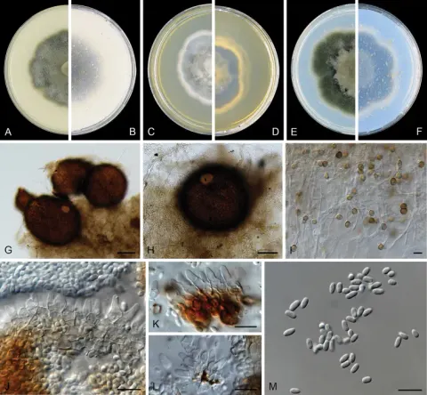
Koloni setelah 7 d di 25 °C, pada OA mencapai 50–55 mm diam, mycelium udara langka, massa spora dengan penampilan seperti lemak, bata gelap ke sepia, kayu manis ke tepi, produksi yang banyak dari pikinidia yang bersempadan, margin…
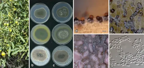
Colonies on OA, 27–30 mm diam. after 7 d, flat with scant aerial mycelia with a few zonate rings, vinaceous to dark vinaceous; vinaceous to dark vinaceous; on MEA, 23–25 mm after 7 d, margin entire, flat, scant aerial mycelium towards…
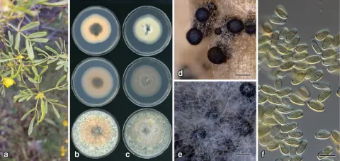
Colonies on OA, 21–25 mm diam. after 7 d, flat with scant aerial mycelia, rosy vinaceous, dark at centre; reverse rosy buff, dark at centre, with a few dark radiating fissures; on MEA, 27–30 mm after 7 d, margin entire, flat, with sparse…