Ciri dan struktur
Stroma solitary, simple, cylindrical, up to 11 cm long, 1 mm wide, brown (oac 48-50), ca. 8.5 cm emerging above the leaf litter, ca. 2.5 cm buried in the soil. Asexual state (Hirsutella) produced ca. 1 cm at the terminal part of the stroma, light brown to grey. Perithecia pseudo-immersed, broadly ovoid covering middle part of stroma, (400–)414–469 (–500) × (200–)208–263(–300) µm. Asci 8-spored, filiform, (187.5–) 217–265(–287.5) × 4.5–6.5(–7.5) µm with cap, 2 µm. Ascospores whole, filiform, (230–)240–291(–315) × 1.5–3 µm, with septa. Colonies on PDA, attaining a diam. of 16 mm within 20 d at 25 °C, cream (oac872) to grey (oac909). Conidiogenous cells swollen towards the base, hyaline, smooth, tapering gradually towards the apex, which often forms a thin neck, monophialidic, (10–)12.5–16 (–17) × (2–) 2.5–3 µm. Conidia arising from phialides at the apex of each neck, oval, 3–4.5(–5) × 2–2.5(–3) µm. Colonies on PSA, attaining a diam. of 17 mm within 20 d at 25 °C, white to grey (oac872). Conidiogenous cells swollen towards the base, hyaline, smooth, tapering gradually towards the apex, which often forms a thin neck, monophialidic, (10–)11.5–15(–17) × (2–)2.5–3.5(–4) µm. Conidia arising from phialides at the apex of each neck, oval, (3–)3.5–5(–5.5) × 2–3 µm. Colonies on SDYA /4, attaining a diam. of 17 mm within 20 d at 25 °C, white to grey (oac802). Conidiogenous cells swollen towards the base, hyaline, smooth, tapering gradually towards the apex, which often forms a thin neck, monophialidic or polyphialidic, (9–)10.5–14.5(–17) × (2–)2.5–3 µm. Conidia arising from phialides at the apex of each neck, oval, (3–)3.5–4.5(–5) × 2–3 µm.
Habitat dan peran ekologis
Thailand, only known from Khao Yai National Park. Parasitic on a pair of termites from a reproductive caste (Order Isoptera: Family Termitidae, Subfamily Macrotermitinae) and these specimens were buried in the soil. The fungus emerged from the segment between the prothorax and mesothorax of one of the termite pairs.
- Habitat atau konteks temuan
- Informasi belum tersedia pada sumber rilis ini.
- Substrat
- Informasi belum tersedia pada sumber rilis ini.
- Peran atau konteks
- Belum dikategorikan pada rilis ini.
Reproduksi
Stroma solitary, simple, cylindrical, up to 11 cm long, 1 mm wide, brown (oac 48-50), ca. 8.5 cm emerging above the leaf litter, ca. 2.5 cm buried in the soil. Asexual state (Hirsutella) produced ca. 1 cm at the terminal part of the stroma, light brown to grey. Perithecia pseudo-immersed, broadly ovoid covering middle part of stroma, (400–)414–469 (–500) × (200–)208–263(–300) µm. Asci 8-spored, filiform, (187.5–) 217–265(–287.5) × 4.5–6.5(–7.5) µm with cap, 2 µm. Ascospores whole, filiform, (230–)240–291(–315) × 1.5–3 µm, with septa. Colonies on PDA, attaining a diam. of 16 mm within 20 d at 25 °C, cream (oac872) to grey (oac909). Conidiogenous cells swollen towards the base, hyaline, smooth, tapering gradually towards the apex, which often forms a thin neck, monophialidic, (10–)12.5–16 (–17) × (2–) 2.5–3 µm.
Catatan identitas dan cakupan
- Nama diterima mengikuti Catalogue of Life Base2026-09-11; uraian mempertahankan konsep dan nama sumber aslinya.
- Preserve original treatment localities, hosts and substrate scope. Materials examined describe documented specimens, not the full global range.
- Impor dan terjemahan otomatis belum diperiksa editorial per profil. Struktur reproduktif yang disebutkan tidak membuktikan seluruh siklus hidup diketahui.
- Pengukuran pada uraian mengikuti spesimen, kultur, dan metode sumber; angka tidak diperluas ke semua populasi.
Catatan distribusi dalam narasi mengikuti sumbernya. Dokumentasi spesimen atau foto mengikuti catatan sumber. Bukti wilayah Indonesia dan penilaian konservasi tidak disimpulkan dari gambar.
Melihat dengan konteks
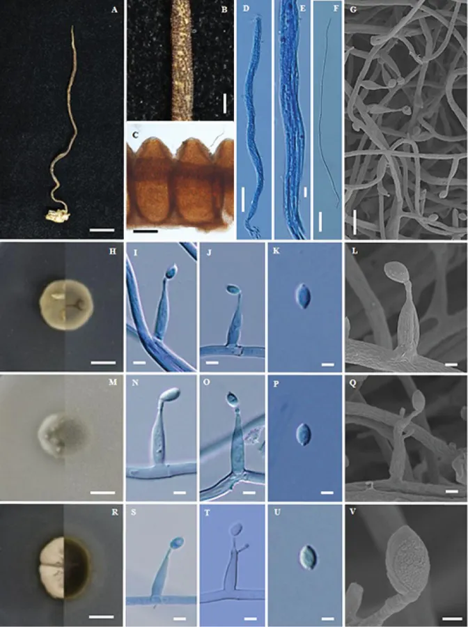
Ophiocordyceps mosingtoensis (BBH26809, BCC36921) A stroma of fungus emerging from termite B part of stroma showing perithecia C pseudo-immersed perithecia D, E ascus F ascospore G, L, Q, V scanning electron micrographs of phialides with conidia on PDA H colony on PDA at 20 d obverse and reverse I, J phialides with conidia K conidium M colony on PSA at 20 d obverse and reverse N, O phialides with conidia P conidium R colony on SDYA /4 at 20 d obverse and reverse S, T phialides with conidia U conidium. Scale bars: 10 mm (A); 1 mm (B); 150 µm (C); 25 µm (D); 4 µm (E); 30 µm (F); 10 µm (G); 8 mm (H, M, R); 3 µm (I, J, N, O, S, T); 2 µm (K, L, P, Q, U); 1 µm (V).
- Jenis
- Pelat ilmiah
- Kredit
- Kanoksri Tasanathai, Wasana Noisripoom, Thanyarat Chaitika, Artit Khonsanit, Sasitorn Hasin, Jennifer Luangsa-ard
- Lisensi
- CC BY 4.0
- Sumber media
- Buka entri sumber
Sumber profil
- [1] Phylogenetic and morphological classification of Ophiocordyceps species on termites from Thailand — Ophiocordyceps mosingtoensis
- [2] Catalogue of Life Base2026-09-11 — Ophiocordyceps mosingtoensis
- [3] Dokumentasi visual — Ophiocordyceps mosingtoensis
Rujukan halaman ini
Fungi. Ophiocordyceps mosingtoensis. Rilis data fungi-2026-10-12-expansion-2. https://fungi-atlas.mgadib.chatgpt.site/jamur/ophiocordyceps-mosingtoensis/.
Status: impor sumber; pemeriksaan editorial per profil belum selesai. Untuk klaim biologis, gunakan rujukan pada bagian terkait dan periksa konteks sumber aslinya.