01 / Morfologi

Ciri dan struktur

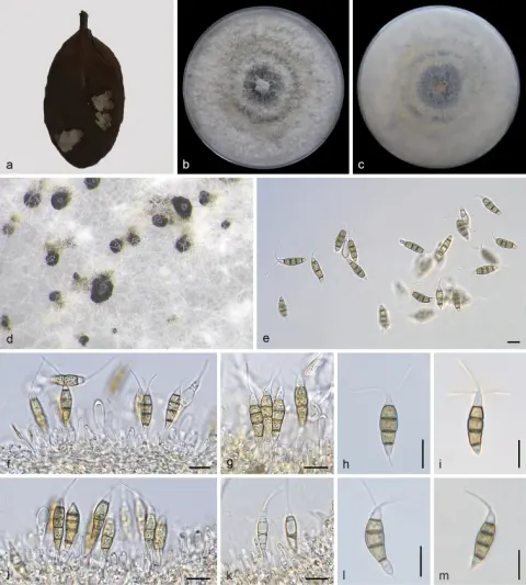
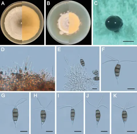
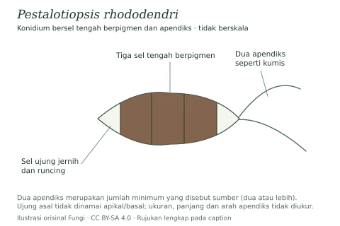
On carnation leaves (Dianthus caryophyllus) supplanted on WA (NTUPPMCC 18-162). Sexual morph was not observed in culture. Asexual morph: Conidiomata acervular, globose, solitary or gregarious, semi-immersed, 100–500 μm diam.; oozing globose to clavate, black conidial masses. Conidiophores subcylindrical, hyaline, smooth, annelidic, indistinct and frequently merged to conidiogenous cells. Conidiogenous cells long pyriform to cylindrical, hyaline, smooth, (1.8–)2.0–3.7(–5.6) × (6.3–)9.7–17.2(–23.2) μm, x– ± SD = 2.9 ± 0.8 × 13.5 ± 3.8 μm. Conidia fusoid, straight or slightly curved, 4-septate, smooth, (4.7–)5.5–6.5(–7.5) × (21.1–)22.4–25.4(–27.4) μm, x– ± SD = 6.0 ± 0.5 × 23.9 ± 1.5 μm, bearing appendages; basal cell obconic with a truncate base, hyaline or pale brown, thin-walled, (0.8–)4.0–6.1(–6.6) μm long, x– ± SD = 5.0 ± 1.0 μm; three median cells long doliiform to subcylindrical, pale brown, concolourous, thick-walled, the first median cell from base (4.1–)4.5–5.3(–5.8) μm long (x– ± SD = 4.9 ± 0.4 μm), the second median cell (3.8–)4.3–5.0(–5.3) μm long (x–± SD = 4.6 ± 0.3 μm), the third median cell (4.3–)4.6–5.4(–6.3) μm long (x– ± SD = 5.0 ± 0.4 μm), together (12.7–)13.7–15.2 (–16.0) μm long (x– ± SD = 14.5 ± 0.8 μm); apical cell conical to subcylindrical with a truncate or acute apex, hyaline, thick-walled, (3.5–)3.8–4.7(–5.2) μm long (x– ± SD = 4.3 ± 0.5 μm). Appendages tubular, hyaline, straight or slightly bent, apical appendage 2–3, unbranched, (8.5–)12.1–18.7(–24.9) μm long (x– ± SD = 15.4 ± 3.3 μm), basal appendage 1–3 (mostly single), centric, unbranched (rarely branched), (2.5–)3.2–6.1(–8.8) μm long (x– ± SD = 4.7 ± 1.4 μm).

Sebagian uraian ditampilkan dalam bahasa sumber; terjemahannya belum tersedia atau belum lolos pemeriksaan.

Rujukan: [1]

Pengukuran mengikuti objek, spesimen atau kultur, dan metode yang disebutkan sumber. Baca uraian asli untuk konteks lengkap.

02 / Kehidupan

Habitat dan peran ekologis

Colonies on PDA reaching 71.9 mm diam. on average after culturing at 25 °C in the dark for seven days, circular, flat with slightly undulate edge, aerial mycelium dense, white; reverse yellowish. Taiwan, Taoyuan City, Dongyanshan, on the stroma of Ophiocordyceps sp. parasitic on an insect (Hymenoptera), 7 October 2018, Wei-Yu Chuang, living culture NTUPPMCC 18-162 (= CD03). Multi-locus phylogenetic analysis indicated that strain NTUPPMCC 18-162 was clustered in the same clade with the ex-type strain of P. hispanica CBS 115391 with absolute statistical support (MLB = 100%, MPB = 100%, PP = 1.00). Even though the DNA sequences of ITS (100%), tub2 (99.22%) and tef1-α (100%) genes of NTUPPMCC 18-162 were very similar to the ex-type strain of P. hispanica (CBS 115391), the morphology of our strain is somewhat different from the original description of P. hispanica (holotype CBS H-23554) published by Liu et al. (2019). For instance, NTUPPMCC 18-162 has longer apical appendages (12–18 μm versus 2–14 μm) and contains higher numbers of basal appendages (0–1 versus 0–3) (Suppl. material 1: table S7) compared to the P. hispanica holotype CBS H-23554. However, P. hispanica was originally isolated from Protea cv. ‘Susara’ (Proteaceae) and collected in Spain, while NTUPPMCC 18-162 was isolated from the stroma of Ophiocordyceps sp. in Taiwan.

Sebagian uraian ditampilkan dalam bahasa sumber; terjemahannya belum tersedia atau belum lolos pemeriksaan.

Rujukan: [1]

Habitat atau konteks temuan
kultur yang didokumentasikan
Substrat
Informasi belum tersedia pada sumber rilis ini.
Peran atau konteks
Belum dikategorikan pada rilis ini.
Temukan hubungan serupa
03 / Siklus hidup

Reproduksi

On carnation leaves (Dianthus caryophyllus) supplanted on WA (NTUPPMCC 18-162). Sexual morph was not observed in culture. Asexual morph: Conidiomata acervular, globose, solitary or gregarious, semi-immersed, 100–500 μm diam.; oozing globose to clavate, black conidial masses. Conidiophores subcylindrical, hyaline, smooth, annelidic, indistinct and frequently merged to conidiogenous cells. Conidiogenous cells long pyriform to cylindrical, hyaline, smooth, (1.8–)2.0–3.7(–5.6) × (6.3–)9.7–17.2(–23.2) μm, x– ± SD = 2.9 ± 0.8 × 13.5 ± 3.8 μm. Conidia fusoid, straight or slightly curved, 4-septate, smooth, (4.7–)5.5–6.5(–7.5) × (21.1–)22.4–25.4(–27.4) μm, x– ± SD = 6.0 ± 0.5 × 23.9 ± 1.5 μm, bearing appendages; basal cell obconic with a truncate base, hyaline or pale brown, thin-walled, (0.8–)4.0–6.1(–6.6) μm long, x– ± SD = 5.0 ± 1.0 μm; three median cells long doliiform to subcylindrical, pale brown, concolourous, thick-walled, the first median cell from base (4.1–)4.5–5.3(–5.8) μm long (x– ± SD = 4.9 ± 0.4 μm), the second median cell (3.8–)4.3–5.0(–5.3) μm long (x–± SD = 4.6 ± 0.3 μm), the third median cell (4.3–)4.6–5.4(–6.3) μm long (x– ± SD = 5.0 ± 0.4 μm), together (12.7–)13.7–15.2 (–16.0) μm long (x– ± SD = 14.5 ± 0.8 μm); apical cell conical to subcylindrical with a truncate or acute apex, hyaline, thick-walled, (3.5–)3.8–4.7(–5.2) μm long (x– ± SD = 4.3 ± 0.5 μm).

Sebagian uraian ditampilkan dalam bahasa sumber; terjemahannya belum tersedia atau belum lolos pemeriksaan.

Rujukan: [1]

04 / Batas informasi

Catatan identitas dan cakupan

  • Nama diterima mengikuti Catalogue of Life Base2026-09-11; uraian mempertahankan konsep dan nama sumber aslinya.
  • Preserve original treatment localities, hosts and substrate scope. Materials examined describe documented specimens, not the full global range.
  • Impor dan terjemahan otomatis belum diperiksa editorial per profil. Struktur reproduktif yang disebutkan tidak membuktikan seluruh siklus hidup diketahui.
  • Pengukuran pada uraian mengikuti spesimen, kultur, dan metode sumber; angka tidak diperluas ke semua populasi.

Catatan distribusi dalam narasi mengikuti sumbernya. Dokumentasi spesimen atau foto mengikuti catatan sumber. Bukti wilayah Indonesia dan penilaian konservasi tidak disimpulkan dari gambar.

05 / Dokumentasi

Melihat dengan konteks

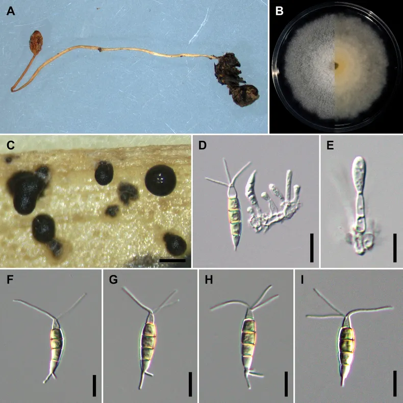
Pestalotiopsis hispanica (NTUPPMCC 18-162 = CD03) A the original habitat of Pestalotiopsis hispanica; the stroma of Ophiocordyceps sp. B top view (left) and bottom view (right) of the colony on potato dextrose agar (PDA) after incubation for seven days C conidiomata on carnation leaf D, E conidiogenous cells and immature conidia F–I conidia. Scale bars: 250 μm (C); 10 μm (D–I).

Jenis
Pelat ilmiah
Kredit
Sheng-Yu Hsu, Yuan-Cheng Xu, Yu-Chen Lin, Wei-Yu Chuang, Shiou-Ruei Lin, Marc Stadler, Narumon Tangthirasunun, Ratchadawan Cheewangkoon, Hind A. AL-Shwaiman, Abdallah M. Elgorban, Hiran A. Ariyawansa
Lisensi
CC BY 4.0
Sumber media
Buka entri sumber

Gambar menunjukkan objek menurut identifikasi sumber, bukan verifikasi independen spesimen oleh Fungi. Tidak ada skala baru yang ditambahkan.

06 / Rujukan

Sumber profil

  1. [1] Hidden diversity of Pestalotiopsis and Neopestalotiopsis (Amphisphaeriales, Sporocadaceae) species allied with the stromata of entomopathogenic fungi in Taiwan — Pestalotiopsis hispanicaBagian sumber: PMC10851163_section_24
    Tanggal penghimpunan sumber: 2026-10-12
    Teks sumber: CC BY 4.0 · Hsu SY, Xu YC, Lin YC, Chuang WY, Lin SR, Stadler M, Tangthirasunun N, Cheewangkoon R, Al-Shwaiman HA, Elgorban AM, Ariyawansa HA.
  2. [2] Catalogue of Life Base2026-09-11 — Pestalotiopsis hispanicaBagian sumber: ColDP NameUsage.tsv ID 4FC3V; accepted species; source 2073
    Tanggal penghimpunan sumber: 2026-10-12
  3. [3] Dokumentasi visual — Pestalotiopsis hispanicaBagian sumber: Individual media record; identification follows the source, not independent determination
    Tanggal penghimpunan sumber: 2026-10-12
    Media sumber: CC BY 4.0 · Sheng-Yu Hsu, Yuan-Cheng Xu, Yu-Chen Lin, Wei-Yu Chuang, Shiou-Ruei Lin, Marc Stadler, Narumon Tangthirasunun, Ratchadawan Cheewangkoon, Hind A. AL-Shwaiman, Abdallah M. Elgorban, Hiran A. Ariyawansa

Adaptasi teks: CC BY 4.0. Hsu SY, Xu YC, Lin YC, Chuang WY, Lin SR, Stadler M, Tangthirasunun N, Cheewangkoon R, Al-Shwaiman HA, Elgorban AM, Ariyawansa HA. — Hidden diversity of Pestalotiopsis and Neopestalotiopsis (Amphisphaeriales, Sporocadaceae) species allied with the stromata of entomopathogenic fungi in Taiwan — Pestalotiopsis hispanica

Uraian asli dan riwayat sumber

Riwayat atau halaman sumber

Ringkasan sumber

On carnation leaves (Dianthus caryophyllus) supplanted on WA (NTUPPMCC 18-162). Sexual morph was not observed in culture. Asexual morph: Conidiomata acervular, globose, solitary or gregarious, semi-immersed, 100–500 μm diam.; oozing globose to clavate, black conidial masses. Conidiophores subcylindrical, hyaline, smooth, annelidic, indistinct and frequently merged to conidiogenous cells. Conidiogenous cells long pyriform to cylindrical, hyaline, smooth, (1.8–)2.0–3.7(–5.6) × (6.3–)9.7–17.2(–23.2) μm, x– ± SD = 2.9 ± 0.8 × 13.5 ± 3.8 μm.

Revisi sumber

Morfologi sumber

On carnation leaves (Dianthus caryophyllus) supplanted on WA (NTUPPMCC 18-162). Sexual morph was not observed in culture. Asexual morph: Conidiomata acervular, globose, solitary or gregarious, semi-immersed, 100–500 μm diam.; oozing globose to clavate, black conidial masses. Conidiophores subcylindrical, hyaline, smooth, annelidic, indistinct and frequently merged to conidiogenous cells. Conidiogenous cells long pyriform to cylindrical, hyaline, smooth, (1.8–)2.0–3.7(–5.6) × (6.3–)9.7–17.2(–23.2) μm, x– ± SD = 2.9 ± 0.8 × 13.5 ± 3.8 μm. Conidia fusoid, straight or slightly curved, 4-septate, smooth, (4.7–)5.5–6.5(–7.5) × (21.1–)22.4–25.4(–27.4) μm, x– ± SD = 6.0 ± 0.5 × 23.9 ± 1.5 μm, bearing appendages; basal cell obconic with a truncate base, hyaline or pale brown, thin-walled, (0.8–)4.0–6.1(–6.6) μm long, x– ± SD = 5.0 ± 1.0 μm; three median cells long doliiform to subcylindrical, pale brown, concolourous, thick-walled, the first median cell from base (4.1–)4.5–5.3(–5.8) μm long (x– ± SD = 4.9 ± 0.4 μm), the second median cell (3.8–)4.3–5.0(–5.3) μm long (x–± SD = 4.6 ± 0.3 μm), the third median cell (4.3–)4.6–5.4(–6.3) μm long (x– ± SD = 5.0 ± 0.4 μm), together (12.7–)13.7–15.2 (–16.0) μm long (x– ± SD = 14.5 ± 0.8 μm); apical cell conical to subcylindrical with a truncate or acute apex, hyaline, thick-walled, (3.5–)3.8–4.7(–5.2) μm long (x– ± SD = 4.3 ± 0.5 μm). Appendages tubular, hyaline, straight or slightly bent, apical appendage 2–3, unbranched, (8.5–)12.1–18.7(–24.9) μm long (x– ± SD = 15.4 ± 3.3 μm), basal appendage 1–3 (mostly single), centric, unbranched (rarely branched), (2.5–)3.2–6.1(–8.8) μm long (x– ± SD = 4.7 ± 1.4 μm).

Revisi sumber

Ekologi sumber

Colonies on PDA reaching 71.9 mm diam. on average after culturing at 25 °C in the dark for seven days, circular, flat with slightly undulate edge, aerial mycelium dense, white; reverse yellowish. Taiwan, Taoyuan City, Dongyanshan, on the stroma of Ophiocordyceps sp. parasitic on an insect (Hymenoptera), 7 October 2018, Wei-Yu Chuang, living culture NTUPPMCC 18-162 (= CD03). Multi-locus phylogenetic analysis indicated that strain NTUPPMCC 18-162 was clustered in the same clade with the ex-type strain of P. hispanica CBS 115391 with absolute statistical support (MLB = 100%, MPB = 100%, PP = 1.00). Even though the DNA sequences of ITS (100%), tub2 (99.22%) and tef1-α (100%) genes of NTUPPMCC 18-162 were very similar to the ex-type strain of P. hispanica (CBS 115391), the morphology of our strain is somewhat different from the original description of P. hispanica (holotype CBS H-23554) published by Liu et al. (2019). For instance, NTUPPMCC 18-162 has longer apical appendages (12–18 μm versus 2–14 μm) and contains higher numbers of basal appendages (0–1 versus 0–3) (Suppl. material 1: table S7) compared to the P. hispanica holotype CBS H-23554. However, P. hispanica was originally isolated from Protea cv. ‘Susara’ (Proteaceae) and collected in Spain, while NTUPPMCC 18-162 was isolated from the stroma of Ophiocordyceps sp. in Taiwan.

Revisi sumber

Struktur reproduksi sumber

On carnation leaves (Dianthus caryophyllus) supplanted on WA (NTUPPMCC 18-162). Sexual morph was not observed in culture. Asexual morph: Conidiomata acervular, globose, solitary or gregarious, semi-immersed, 100–500 μm diam.; oozing globose to clavate, black conidial masses. Conidiophores subcylindrical, hyaline, smooth, annelidic, indistinct and frequently merged to conidiogenous cells. Conidiogenous cells long pyriform to cylindrical, hyaline, smooth, (1.8–)2.0–3.7(–5.6) × (6.3–)9.7–17.2(–23.2) μm, x– ± SD = 2.9 ± 0.8 × 13.5 ± 3.8 μm. Conidia fusoid, straight or slightly curved, 4-septate, smooth, (4.7–)5.5–6.5(–7.5) × (21.1–)22.4–25.4(–27.4) μm, x– ± SD = 6.0 ± 0.5 × 23.9 ± 1.5 μm, bearing appendages; basal cell obconic with a truncate base, hyaline or pale brown, thin-walled, (0.8–)4.0–6.1(–6.6) μm long, x– ± SD = 5.0 ± 1.0 μm; three median cells long doliiform to subcylindrical, pale brown, concolourous, thick-walled, the first median cell from base (4.1–)4.5–5.3(–5.8) μm long (x– ± SD = 4.9 ± 0.4 μm), the second median cell (3.8–)4.3–5.0(–5.3) μm long (x–± SD = 4.6 ± 0.3 μm), the third median cell (4.3–)4.6–5.4(–6.3) μm long (x– ± SD = 5.0 ± 0.4 μm), together (12.7–)13.7–15.2 (–16.0) μm long (x– ± SD = 14.5 ± 0.8 μm); apical cell conical to subcylindrical with a truncate or acute apex, hyaline, thick-walled, (3.5–)3.8–4.7(–5.2) μm long (x– ± SD = 4.3 ± 0.5 μm).

Revisi sumber

Rujukan halaman ini

Fungi. Pestalotiopsis hispanica. Rilis data fungi-2026-10-12-expansion-2. https://fungi-atlas.mgadib.chatgpt.site/jamur/pestalotiopsis-hispanica/.

Status: impor sumber; pemeriksaan editorial per profil belum selesai. Untuk klaim biologis, gunakan rujukan pada bagian terkait dan periksa konteks sumber aslinya.