01 / Morfologi

Ciri dan struktur

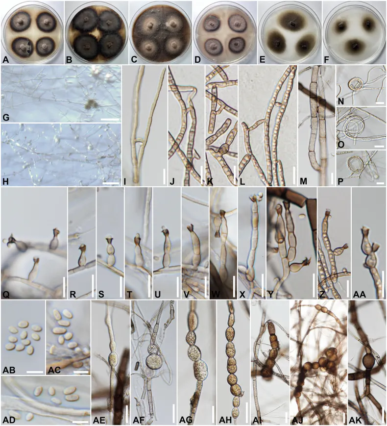
Colonies on different agar media were incubated in the dark at 25 °C for 1 months; colonies flat, irregular, with edge entire, velvety; on PDA (39 to 44 mm in diameter) surface grayish brown, dark brown to black at the margin, reverse olivaceous black; on MEA surface Blackish Grey, black at the margin, reverse olivaceous black; on MEA (49 to 53 mm in diameter) surface Blackish Grey, black at the margin, reverse olivaceous black; on OA (69 to 74 mm in diameter) surface grayish brown, reverse olivaceous black; on PCA (46 to 55 mm in diameter) surface and reverse grayish brown, dark brown at the margin, reverse olivaceous black, sporulation absent; on CMD (42 to 50 mm in diameter) surface and reverse brownish gray; and on CMA (33 to 41 mm in diameter) surface and reverse brownish grey, sporulation absent. Asexual morph in vitro dematiaceous hyphomycetes. Hyphae 1–3.4 µm wide, subhyaline to light brown, simple to branched, septate, smooth-, thin-walled, coiling, anastomosis observed. Conidiophores (4–)5.5–31(–45.3) × (2–)3–4.3(–4.8) µm (x̄ = 17 × 3.6, n = 55), micro- or semi-macronematous, straight, simples or poorly branched, septate, slightly constricted at septa, smooth; micronematous conidiophores consisting in conidiogenous cells growing directly from vegetative hyphae, lateral or terminal. Phialides 4–11(–15.5) × 2.3–4(–5) µm (x̄ = 8.3 × 3.3, n = 75), regularly flask-shaped to elongate-ampulliform or subulate, with an apical conspicuous collarette; collarettes (1.6–)2–3.9(–4.8) × (1.6–)2–3.9(–4.2) µm (x̄ = 3 × 3, n = 65), usually funnel-shaped, darker than the rest of the phialide.

Sebagian uraian ditampilkan dalam bahasa sumber; terjemahannya belum tersedia atau belum lolos pemeriksaan.

Rujukan: [1]

Pengukuran mengikuti objek, spesimen atau kultur, dan metode yang disebutkan sumber. Baca uraian asli untuk konteks lengkap.

02 / Kehidupan

Habitat dan peran ekologis

Endolichenic fungi from the medulla of foliose lichen (Parmotrema sp.) in Thailand (this study); pathogenic as chromoblastomycosis in human in China and Mexico (Li et al. 2017; Ahmed et al. 2021), as fungal keratitis in India and USA (Ply et al. 2023; Mitra et al. 2024), as phaeohyphomycosis in France (Pruvot et al. 2023); saprobic from bamboo in China, from the environment in Japan, from plant materials in Brazil and China, and soil in China and from wheat straw in Brazil (Li et al. 2017).

Sebagian uraian ditampilkan dalam bahasa sumber; terjemahannya belum tersedia atau belum lolos pemeriksaan.

Rujukan: [1]

Habitat atau konteks temuan
Informasi belum tersedia pada sumber rilis ini.
Substrat
Informasi belum tersedia pada sumber rilis ini.
Peran atau konteks
Pengurai
Temukan hubungan serupa
03 / Siklus hidup

Reproduksi

Colonies on different agar media were incubated in the dark at 25 °C for 1 months; colonies flat, irregular, with edge entire, velvety; on PDA (39 to 44 mm in diameter) surface grayish brown, dark brown to black at the margin, reverse olivaceous black; on MEA surface Blackish Grey, black at the margin, reverse olivaceous black; on MEA (49 to 53 mm in diameter) surface Blackish Grey, black at the margin, reverse olivaceous black; on OA (69 to 74 mm in diameter) surface grayish brown, reverse olivaceous black; on PCA (46 to 55 mm in diameter) surface and reverse grayish brown, dark brown at the margin, reverse olivaceous black, sporulation absent; on CMD (42 to 50 mm in diameter) surface and reverse brownish gray; and on CMA (33 to 41 mm in diameter) surface and reverse brownish grey, sporulation absent.

Sebagian uraian ditampilkan dalam bahasa sumber; terjemahannya belum tersedia atau belum lolos pemeriksaan.

Rujukan: [1]

04 / Batas informasi

Catatan identitas dan cakupan

  • Nama diterima mengikuti Catalogue of Life Base2026-09-11; uraian mempertahankan konsep dan nama sumber aslinya.
  • Preserve original treatment localities, hosts and substrate scope. Materials examined describe documented specimens, not the full global range.
  • Impor dan terjemahan otomatis belum diperiksa editorial per profil. Struktur reproduktif yang disebutkan tidak membuktikan seluruh siklus hidup diketahui.
  • Pengukuran pada uraian mengikuti spesimen, kultur, dan metode sumber; angka tidak diperluas ke semua populasi.

Catatan distribusi dalam narasi mengikuti sumbernya. Dokumentasi spesimen atau foto mengikuti catatan sumber. Bukti wilayah Indonesia dan penilaian konservasi tidak disimpulkan dari gambar.

05 / Dokumentasi

Melihat dengan konteks

Phialophora chinensis (SDBR-CMU506, new host record). A–F. Colonies on PDA, MEA, OA, PCA, CMD and CMA, respectively, after 30 d at 25 °C; G. Conidiophores sporulating on CMA; H. Conidiophores sporulating on PCA; I–M. Hyphal anastomosis; N–P. Hyphal coil; Q–AA. Phialides at different stages of development; AB–AD. Conidia; AE–AK. Chlamydospores (intercalary and chains formation). Scale bars: 100 µm (G, H); 10 µm (I–AA); 5 µm (AB–AD); 20 µm (AE–AK).

Jenis
Pelat ilmiah
Kredit
Chanokned Senwanna, Jaturong Kumla, Pratthana Kodchasee, Nutchanan Duangkon, Nakarin Suwannarach
Lisensi
CC BY 4.0
Sumber media
Buka entri sumber

Gambar menunjukkan objek menurut identifikasi sumber, bukan verifikasi independen spesimen oleh Fungi. Tidak ada skala baru yang ditambahkan.

06 / Rujukan

Sumber profil

  1. [1] Additions of New Endolichenic Fungi to Herpotrichiellaceae (Chaetothyriales, Ascomycota) from northern Thailand — Phialophora chinensisBagian sumber: PMC12329419_section_19
    Tanggal penghimpunan sumber: 2026-10-12
    Teks sumber: CC BY 4.0 · Senwanna C, Kumla J, Kodchasee P, Duangkon N, Suwannarach N.
  2. [2] Catalogue of Life Base2026-09-11 — Phialophora chinensisBagian sumber: ColDP NameUsage.tsv ID 4G452; accepted species; source 2073
    Tanggal penghimpunan sumber: 2026-10-12
  3. [3] Dokumentasi visual — Phialophora chinensisBagian sumber: Individual media record; identification follows the source, not independent determination
    Tanggal penghimpunan sumber: 2026-10-12
    Media sumber: CC BY 4.0 · Chanokned Senwanna, Jaturong Kumla, Pratthana Kodchasee, Nutchanan Duangkon, Nakarin Suwannarach

Adaptasi teks: CC BY 4.0. Senwanna C, Kumla J, Kodchasee P, Duangkon N, Suwannarach N. — Additions of New Endolichenic Fungi to Herpotrichiellaceae (Chaetothyriales, Ascomycota) from northern Thailand — Phialophora chinensis

Uraian asli dan riwayat sumber

Riwayat atau halaman sumber

Ringkasan sumber

Colonies on different agar media were incubated in the dark at 25 °C for 1 months; colonies flat, irregular, with edge entire, velvety; on PDA (39 to 44 mm in diameter) surface grayish brown, dark brown to black at the margin, reverse olivaceous black; on MEA surface Blackish Grey, black at the margin, reverse olivaceous black; on MEA (49 to 53 mm in diameter) surface Blackish Grey, black at the margin, reverse olivaceous black; on OA (69 to 74 mm in diameter) surface grayish brown, reverse olivaceous black; on PCA (46 to 55 mm in diameter) surface and reverse grayish brown, dark brown at the margin, reverse olivaceous black, sporulation absent; on CMD (42 to 50 mm in diameter) surface and reverse brownish gray; and on CMA (33 to 41 mm in diameter) surface and reverse brownish grey, sporulation absent.

Revisi sumber

Morfologi sumber

Colonies on different agar media were incubated in the dark at 25 °C for 1 months; colonies flat, irregular, with edge entire, velvety; on PDA (39 to 44 mm in diameter) surface grayish brown, dark brown to black at the margin, reverse olivaceous black; on MEA surface Blackish Grey, black at the margin, reverse olivaceous black; on MEA (49 to 53 mm in diameter) surface Blackish Grey, black at the margin, reverse olivaceous black; on OA (69 to 74 mm in diameter) surface grayish brown, reverse olivaceous black; on PCA (46 to 55 mm in diameter) surface and reverse grayish brown, dark brown at the margin, reverse olivaceous black, sporulation absent; on CMD (42 to 50 mm in diameter) surface and reverse brownish gray; and on CMA (33 to 41 mm in diameter) surface and reverse brownish grey, sporulation absent. Asexual morph in vitro dematiaceous hyphomycetes. Hyphae 1–3.4 µm wide, subhyaline to light brown, simple to branched, septate, smooth-, thin-walled, coiling, anastomosis observed. Conidiophores (4–)5.5–31(–45.3) × (2–)3–4.3(–4.8) µm (x̄ = 17 × 3.6, n = 55), micro- or semi-macronematous, straight, simples or poorly branched, septate, slightly constricted at septa, smooth; micronematous conidiophores consisting in conidiogenous cells growing directly from vegetative hyphae, lateral or terminal. Phialides 4–11(–15.5) × 2.3–4(–5) µm (x̄ = 8.3 × 3.3, n = 75), regularly flask-shaped to elongate-ampulliform or subulate, with an apical conspicuous collarette; collarettes (1.6–)2–3.9(–4.8) × (1.6–)2–3.9(–4.2) µm (x̄ = 3 × 3, n = 65), usually funnel-shaped, darker than the rest of the phialide.

Revisi sumber

Ekologi sumber

Endolichenic fungi from the medulla of foliose lichen (Parmotrema sp.) in Thailand (this study); pathogenic as chromoblastomycosis in human in China and Mexico (Li et al. 2017; Ahmed et al. 2021), as fungal keratitis in India and USA (Ply et al. 2023; Mitra et al. 2024), as phaeohyphomycosis in France (Pruvot et al. 2023); saprobic from bamboo in China, from the environment in Japan, from plant materials in Brazil and China, and soil in China and from wheat straw in Brazil (Li et al. 2017).

Revisi sumber

Struktur reproduksi sumber

Colonies on different agar media were incubated in the dark at 25 °C for 1 months; colonies flat, irregular, with edge entire, velvety; on PDA (39 to 44 mm in diameter) surface grayish brown, dark brown to black at the margin, reverse olivaceous black; on MEA surface Blackish Grey, black at the margin, reverse olivaceous black; on MEA (49 to 53 mm in diameter) surface Blackish Grey, black at the margin, reverse olivaceous black; on OA (69 to 74 mm in diameter) surface grayish brown, reverse olivaceous black; on PCA (46 to 55 mm in diameter) surface and reverse grayish brown, dark brown at the margin, reverse olivaceous black, sporulation absent; on CMD (42 to 50 mm in diameter) surface and reverse brownish gray; and on CMA (33 to 41 mm in diameter) surface and reverse brownish grey, sporulation absent.

Revisi sumber

Rujukan halaman ini

Fungi. Phialophora chinensis. Rilis data fungi-2026-10-12-expansion-2. https://fungi-atlas.mgadib.chatgpt.site/jamur/phialophora-chinensis/.

Status: impor sumber; pemeriksaan editorial per profil belum selesai. Untuk klaim biologis, gunakan rujukan pada bagian terkait dan periksa konteks sumber aslinya.
