01 / Morfologi

Ciri dan struktur

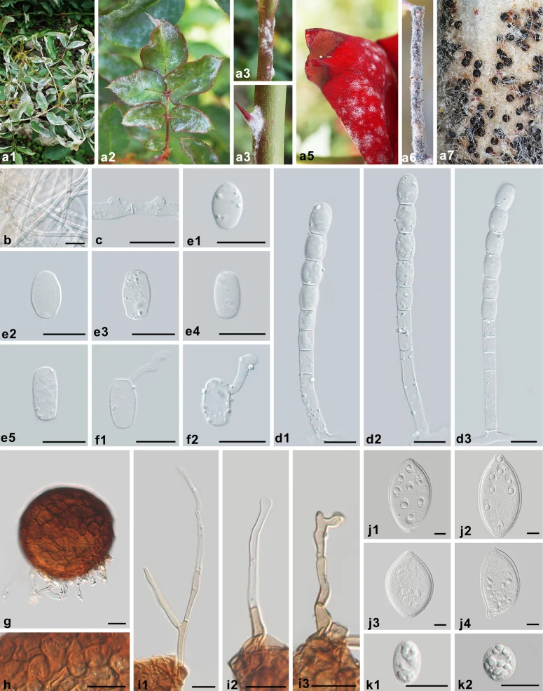
Mycelium on leaves, amphigenous, also on stems and petals, persistent, infected shoots often curled and distorted, forming irregular patches or effuse; primary hyphae hyaline, thin-walled, straight or sometimes flexuous, (2.5–)3–5.5 μm wide; secondary hyphae hyaline, thick-walled, 4–6 μm wide, colonies on twigs only developed in the later stages of the disease; hyphal appressoria indistinct to nipple-shaped, solitary or side by side; conidiophores arising from the upper surface of mother cells, erect, (52–)60–97(–99) × (6.5–)7.5–9.5 μm, foot cells straight, cylindrical, 33–59 × 7–9.5 μm, followed by (1–)2–3 shorter cells, forming catenescent conidia; conidia variable, primary conidia obovoid, secondary conidia doliiform to cylindrical, 20–26 × 11.5–15 μm, length/width ratio 1.5–2.1; germ tubes almost terminal. Chasmothecia gregarious, subglobose to globose, immersed in the secondary mycelial patches, mostly on stems, but also produced on leaves when the disease is severe, (74–)88–113 μm diam.; peridium cells round to irregularly polygonal, 4.0–16 μm diam.; appendages few, in the lower half of chasmothecia, mycelioid, short, usually shorter than chasmothecial diam., rarely up to 1.5 times diam., occasionally branching, brownish at the basal part or brown throughout, septate, thin-walled, smooth, coarse; ascus ovoid to fusiform, colorless, thick-walled, sessile or almost sessile, 87–140 × 63–84 μm, length/width ratio 1.2–1.9, terminal oculus obvious, large, 13.5–21 μm, 8-spored; ascospores ellipsoid to round, colorless, (16.5–)18–25 × 12.5–17.5 μm, length/width ratio 1.1–1.9.

Sebagian uraian ditampilkan dalam bahasa sumber; terjemahannya belum tersedia atau belum lolos pemeriksaan.

Rujukan: [1]

Pengukuran mengikuti objek, spesimen atau kultur, dan metode yang disebutkan sumber. Baca uraian asli untuk konteks lengkap.

02 / Kehidupan

Habitat dan peran ekologis

(phylogenetically confirmed hosts and distribution). Rosa (canina, chinensis, × damascena, foetida, gallica, laxa, majalis, multiflora, albertiirubiginosa, rugosa, xanthina), Prunus (armeniaca, cerasus, laurocerasus, persica, granatum), Rosaceae; Catharanthus roseus, Apocynaceae; Citrus reticulata × C. sinensis, Rutaceae; Corymbia citriodora, Eucalyptus (benthamii, grandis, sp.), Myrtaceae; and Forsythia × intermedia, Oleaceae. Asia (China, India, Iraq, Israel, Japan, Pakistan, South Korea, Vietnam), Europe (Belgium, France, Germany, Netherlands, Serbia, Spain, United Kingdom), North America (Mexico, United States), Oceania (Australia, New Zealand), and South America (Argentina, Brazil, Chile, Peru, Uruguay).

Sebagian uraian ditampilkan dalam bahasa sumber; terjemahannya belum tersedia atau belum lolos pemeriksaan.

Rujukan: [1]

Habitat atau konteks temuan
Informasi belum tersedia pada sumber rilis ini.
Substrat
Informasi belum tersedia pada sumber rilis ini.
Peran atau konteks
Belum dikategorikan pada rilis ini.
Temukan hubungan serupa
03 / Siklus hidup

Reproduksi

Mycelium on leaves, amphigenous, also on stems and petals, persistent, infected shoots often curled and distorted, forming irregular patches or effuse; primary hyphae hyaline, thin-walled, straight or sometimes flexuous, (2.5–)3–5.5 μm wide; secondary hyphae hyaline, thick-walled, 4–6 μm wide, colonies on twigs only developed in the later stages of the disease; hyphal appressoria indistinct to nipple-shaped, solitary or side by side; conidiophores arising from the upper surface of mother cells, erect, (52–)60–97(–99) × (6.5–)7.5–9.5 μm, foot cells straight, cylindrical, 33–59 × 7–9.5 μm, followed by (1–)2–3 shorter cells, forming catenescent conidia; conidia variable, primary conidia obovoid, secondary conidia doliiform to cylindrical, 20–26 × 11.5–15 μm, length/width ratio 1.5–2.1; germ tubes almost terminal.

Sebagian uraian ditampilkan dalam bahasa sumber; terjemahannya belum tersedia atau belum lolos pemeriksaan.

Rujukan: [1]

Sinonim menurut acuan taksonomi rilis: Acrosporium leucoconium; Albigo pannosa; Alphitomorpha pannosa; Crocysporium leuconium; Erysiphe pannosa; Leucothallia pannosa; Oidium forsythiae; Oidium leucoconium; Oidium leuconium; Sphaerotheca macularis f. rosae; Sphaerotheca pannosa; Sphaerotheca pannosa var. persicae; Sphaerotheca pannosa var. rosae; Sphaerotheca persicae; Sphaerotheca rosae
04 / Batas informasi

Catatan identitas dan cakupan

  • Nama diterima mengikuti Catalogue of Life Base2026-09-11; uraian mempertahankan konsep dan nama sumber aslinya.
  • Preserve original treatment localities, hosts and substrate scope. Materials examined describe documented specimens, not the full global range.
  • Impor dan terjemahan otomatis belum diperiksa editorial per profil. Struktur reproduktif yang disebutkan tidak membuktikan seluruh siklus hidup diketahui.
  • Pengukuran pada uraian mengikuti spesimen, kultur, dan metode sumber; angka tidak diperluas ke semua populasi.

Catatan distribusi dalam narasi mengikuti sumbernya. Dokumentasi spesimen atau foto mengikuti catatan sumber. Bukti wilayah Indonesia dan penilaian konservasi tidak disimpulkan dari gambar.

05 / Dokumentasi

Melihat dengan konteks

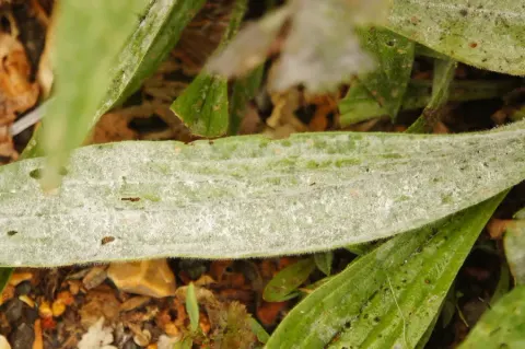
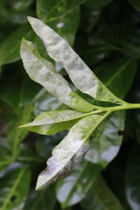
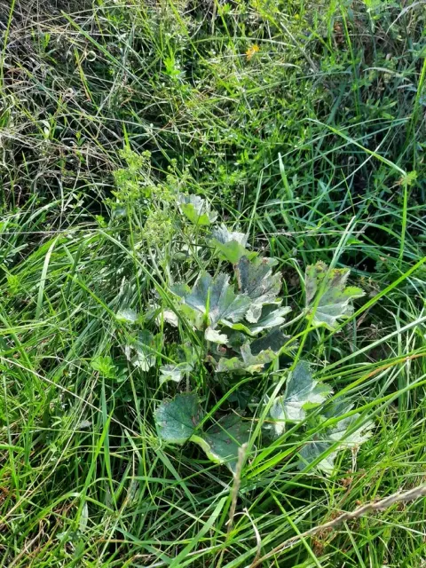
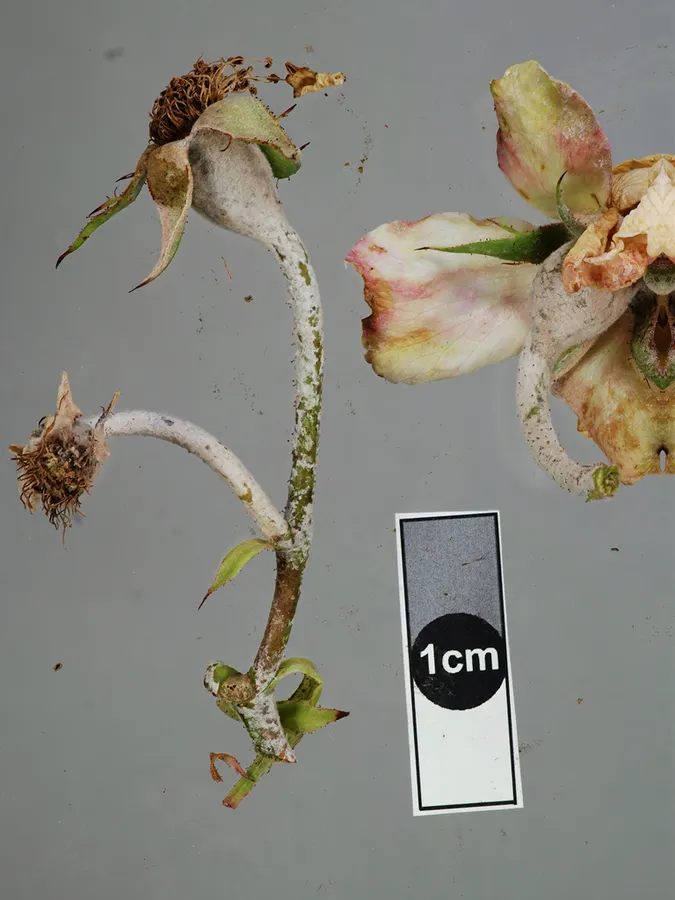
Dokumentasi Podosphaera pannosa menurut asosiasi foto pada record takson iNaturalist. Kredit: Jerry Cooper. Identitas mengikuti sumber; bukan penentuan spesimen independen.

Jenis
Fotografi
Kredit
(c) Jerry Cooper, some rights reserved (CC BY), uploaded by Jerry Cooper
Lisensi
CC BY 4.0
Sumber media
Buka entri sumber

Gambar menunjukkan objek menurut identifikasi sumber, bukan verifikasi independen spesimen oleh Fungi. Tidak ada skala baru yang ditambahkan.

Dokumentasi tambahan

06 / Rujukan

Sumber profil

  1. [1] Revision of powdery mildews (Ascomycota, Erysiphaceae) on Rosa in China: unexpected taxonomic complexity with phytopathological implications — Podosphaera pannosaBagian sumber: PMC12980140_section_23
    Tanggal penghimpunan sumber: 2026-10-12
    Teks sumber: CC BY 4.0 · Jin DN, Wang SB, Guan LP, Wu XL, Feng J, Liu L, Bradshaw M, Braun U, Yang JH, Yu SR, Li Y, Liu SY.
  2. [2] Catalogue of Life Base2026-09-11 — Podosphaera pannosaBagian sumber: ColDP NameUsage.tsv ID 4KRHT; accepted species; source 2073
    Tanggal penghimpunan sumber: 2026-10-12
  3. [3] Dokumentasi visual — Podosphaera pannosaBagian sumber: Identitas mengikuti sumber dokumentasi; bukan penentuan spesimen independen.
    Tanggal penghimpunan sumber: 2026-10-17
    Media sumber: CC BY 4.0 · (c) Jerry Cooper, some rights reserved (CC BY), uploaded by Jerry Cooper
  4. [4] Dokumentasi visual — Podosphaera pannosaBagian sumber: Identitas mengikuti sumber dokumentasi; bukan penentuan spesimen independen.
    Tanggal penghimpunan sumber: 2026-10-17
    Teks sumber: CC BY 4.0 · Dan-Ni Jin, Shuang-Bao Wang, Le-Ping Guan, Xue-Lian Wu, Jing Feng, Li Liu, Michael Bradshaw, Uwe Braun, Jing-Han Yang, Shou-Rong Yu, Yu Li, Shu-Yan Liu

Adaptasi teks: CC BY 4.0. Jin DN, Wang SB, Guan LP, Wu XL, Feng J, Liu L, Bradshaw M, Braun U, Yang JH, Yu SR, Li Y, Liu SY. — Revision of powdery mildews (Ascomycota, Erysiphaceae) on Rosa in China: unexpected taxonomic complexity with phytopathological implications — Podosphaera pannosa

Uraian asli dan riwayat sumber

Riwayat atau halaman sumber

Ringkasan sumber

Mycelium on leaves, amphigenous, also on stems and petals, persistent, infected shoots often curled and distorted, forming irregular patches or effuse; primary hyphae hyaline, thin-walled, straight or sometimes flexuous, (2.5–)3–5.5 μm wide; secondary hyphae hyaline, thick-walled, 4–6 μm wide, colonies on twigs only developed in the later stages of the disease; hyphal appressoria indistinct to nipple-shaped, solitary or side by side; conidiophores arising from the upper surface of mother cells, erect, (52–)60–97(–99) × (6.5–)7.5–9.5 μm, foot cells straight, cylindrical, 33–59 × 7–9.5 μm, followed by (1–)2–3 shorter cells, forming catenescent conidia; conidia variable, primary conidia obovoid, secondary conidia doliiform to cylindrical, 20–26 × 11.5–15 μm, length/width ratio 1.5–2.1; germ tubes almost terminal.

Revisi sumber

Morfologi sumber

Mycelium on leaves, amphigenous, also on stems and petals, persistent, infected shoots often curled and distorted, forming irregular patches or effuse; primary hyphae hyaline, thin-walled, straight or sometimes flexuous, (2.5–)3–5.5 μm wide; secondary hyphae hyaline, thick-walled, 4–6 μm wide, colonies on twigs only developed in the later stages of the disease; hyphal appressoria indistinct to nipple-shaped, solitary or side by side; conidiophores arising from the upper surface of mother cells, erect, (52–)60–97(–99) × (6.5–)7.5–9.5 μm, foot cells straight, cylindrical, 33–59 × 7–9.5 μm, followed by (1–)2–3 shorter cells, forming catenescent conidia; conidia variable, primary conidia obovoid, secondary conidia doliiform to cylindrical, 20–26 × 11.5–15 μm, length/width ratio 1.5–2.1; germ tubes almost terminal. Chasmothecia gregarious, subglobose to globose, immersed in the secondary mycelial patches, mostly on stems, but also produced on leaves when the disease is severe, (74–)88–113 μm diam.; peridium cells round to irregularly polygonal, 4.0–16 μm diam.; appendages few, in the lower half of chasmothecia, mycelioid, short, usually shorter than chasmothecial diam., rarely up to 1.5 times diam., occasionally branching, brownish at the basal part or brown throughout, septate, thin-walled, smooth, coarse; ascus ovoid to fusiform, colorless, thick-walled, sessile or almost sessile, 87–140 × 63–84 μm, length/width ratio 1.2–1.9, terminal oculus obvious, large, 13.5–21 μm, 8-spored; ascospores ellipsoid to round, colorless, (16.5–)18–25 × 12.5–17.5 μm, length/width ratio 1.1–1.9.

Revisi sumber

Ekologi sumber

(phylogenetically confirmed hosts and distribution). Rosa (canina, chinensis, × damascena, foetida, gallica, laxa, majalis, multiflora, albertiirubiginosa, rugosa, xanthina), Prunus (armeniaca, cerasus, laurocerasus, persica, granatum), Rosaceae; Catharanthus roseus, Apocynaceae; Citrus reticulata × C. sinensis, Rutaceae; Corymbia citriodora, Eucalyptus (benthamii, grandis, sp.), Myrtaceae; and Forsythia × intermedia, Oleaceae. Asia (China, India, Iraq, Israel, Japan, Pakistan, South Korea, Vietnam), Europe (Belgium, France, Germany, Netherlands, Serbia, Spain, United Kingdom), North America (Mexico, United States), Oceania (Australia, New Zealand), and South America (Argentina, Brazil, Chile, Peru, Uruguay).

Revisi sumber

Struktur reproduksi sumber

Mycelium on leaves, amphigenous, also on stems and petals, persistent, infected shoots often curled and distorted, forming irregular patches or effuse; primary hyphae hyaline, thin-walled, straight or sometimes flexuous, (2.5–)3–5.5 μm wide; secondary hyphae hyaline, thick-walled, 4–6 μm wide, colonies on twigs only developed in the later stages of the disease; hyphal appressoria indistinct to nipple-shaped, solitary or side by side; conidiophores arising from the upper surface of mother cells, erect, (52–)60–97(–99) × (6.5–)7.5–9.5 μm, foot cells straight, cylindrical, 33–59 × 7–9.5 μm, followed by (1–)2–3 shorter cells, forming catenescent conidia; conidia variable, primary conidia obovoid, secondary conidia doliiform to cylindrical, 20–26 × 11.5–15 μm, length/width ratio 1.5–2.1; germ tubes almost terminal.

Revisi sumber

Rujukan halaman ini

Fungi. Podosphaera pannosa. Rilis data fungi-2026-10-12-expansion-2. https://fungi-atlas.mgadib.chatgpt.site/jamur/podosphaera-pannosa/.

Status: impor sumber; pemeriksaan editorial per profil belum selesai. Untuk klaim biologis, gunakan rujukan pada bagian terkait dan periksa konteks sumber aslinya.