Profil ini diimpor dari uraian spesies pada sumber berlisensi dan sebagian teks diterjemahkan otomatis. Pemeriksaan editorial per profil belum selesai. Bagian yang belum diterjemahkan atau belum lolos pemeriksaan terjemahan tetap ditampilkan dalam bahasa sumber. Angka dan nama ilmiah dipertahankan.
Saprobic on fallen dead leaves. Sexual morph Undetermined. Asexual morph Synnemata 100–130 μm high, 5.9–8 μm wide, erect, simple, unbranched, dark brown to black, comprising various conidiophores twisted together in synnemata, with bubble-like, tightly interwoven, branched hyphae, compacted into an elongate bundle. Conidiophores 100–130 μm long × 1.8–3.5 μm diam. (\(\bar{x} \) = 124 × 2.6 μm, n = 10). Conidiogenous cells 2.5–3 μm long × 1.6–2 μm diam. (\(\bar{x} \) = 2.8 × 1.8 μm, n = 20), monoblastic, integrated, terminal, determinate or percurrent, cylindrical, doliiform, hyaline, smooth, thin-walled. Conidia 13–28 μm long × (2.5–)2.7–3.6(–4) μm diam. (\(\bar{x} \) = 21 × 3.1 μm, n = 20), acrogenous, solitary, hyaline, ellipsoidal, clavate, 2–4-septate, dry, simple, straight, curved, fusiform, smooth- and thin-walled. Culture characteristics: Conidia germinating on PDA within 14 h and germ tubes produced from top cells. Colonies growing on PDA, hairy or cottony, grey to dark grey, reaching 5 mm in 20 days at 25 °C, mycelium superficial, effuse, grey to dark grey hyphae; Asexual spores and sexual spores were not formed within 60 days.
Sebagian uraian ditampilkan dalam bahasa sumber; terjemahannya belum tersedia atau belum lolos pemeriksaan.
Pengukuran mengikuti objek, spesimen atau kultur, dan metode yang disebutkan sumber. Baca uraian asli untuk konteks lengkap.
02 / Kehidupan
Habitat dan peran ekologis
Saprobic on fallen dead leaves. Sexual morph Undetermined. Asexual morph Synnemata 100–130 μm high, 5.9–8 μm wide, erect, simple, unbranched, dark brown to black, comprising various conidiophores twisted together in synnemata, with bubble-like, tightly interwoven, branched hyphae, compacted into an elongate bundle. Conidiophores 100–130 μm long × 1.8–3.5 μm diam. (\(\bar{x} \) = 124 × 2.6 μm, n = 10). Conidiogenous cells 2.5–3 μm long × 1.6–2 μm diam. (\(\bar{x} \) = 2.8 × 1.8 μm, n = 20), monoblastic, integrated, terminal, determinate or percurrent, cylindrical, doliiform, hyaline, smooth, thin-walled. Conidia 13–28 μm long × (2.5–)2.7–3.6(–4) μm diam. (\(\bar{x} \) = 21 × 3.1 μm, n = 20), acrogenous, solitary, hyaline, ellipsoidal, clavate, 2–4-septate, dry, simple, straight, curved, fusiform, smooth- and thin-walled. Culture characteristics: Conidia germinating on PDA within 14 h and germ tubes produced from top cells. Colonies growing on PDA, hairy or cottony, grey to dark grey, reaching 5 mm in 20 days at 25 °C, mycelium superficial, effuse, grey to dark grey hyphae; Asexual spores and sexual spores were not formed within 60 days.
Sebagian uraian ditampilkan dalam bahasa sumber; terjemahannya belum tersedia atau belum lolos pemeriksaan.
Nama diterima mengikuti Catalogue of Life Base2026-09-11; uraian mempertahankan konsep dan nama sumber aslinya.
Preserve original monograph treatment localities, hosts and specimen/substrate scope. Materials examined document specimens, not the full global range.
Impor dan terjemahan otomatis belum diperiksa editorial per profil. Struktur reproduktif yang disebutkan tidak membuktikan seluruh siklus hidup diketahui.
Pengukuran pada uraian mengikuti spesimen, kultur, dan metode sumber; angka tidak diperluas ke semua populasi.
Catatan distribusi dalam narasi mengikuti sumbernya. Dokumentasi spesimen atau foto mengikuti catatan sumber. Bukti wilayah Indonesia dan penilaian konservasi tidak disimpulkan dari gambar.
05 / Dokumentasi
Melihat dengan konteks
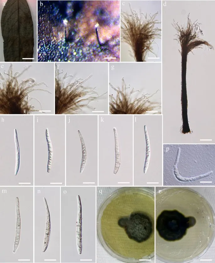
Pseudocercospora maetaengensis (MFLU 14-0206, holotype). a, b Appearance of fungal colonies on host substrate. c, e – g Close up conidiophores and conidiogenous cells. d Conidiophores. h – o Conidia. p Germination of conidia. q – r Culture. Scale bars: a = 1 cm, b = 50 µm, c – d, e – g = 10 µm, h – p = 5 µm, q – r = 0.5 cm
Jenis
Pelat ilmiah
Kredit
Penulis dan sumber artikel: Hyde, Kevin D.; Dong, Yang; Phookamsak, Rungtiwa; Jeewon, Rajesh; Bhat, D. Jayarama; Jones, E. B. Gareth; Liu, Ning-Guo; Abeywickrama, Pranami D.; Mapook, Ausana; Wei, Deping; Perera, Rekhani H.; Manawasinghe, Ishara S.; Pem, Dhandevi; Bundhun, Digvijayini; Karunarathna, Anuruddha; Ekanayaka, Anusha H.; Bao, Dan-Feng; Li, Junfu; Samarakoon, Milan C.; Chaiwan, Napalai; Lin, Chuan-Gen; Phutthacharoen, Kunthida; Zhang, Sheng-Nan; Senanayake, Indunil C.; Goonasekara, Ishani D.; Thambugala, Kasun M.; Phukhamsakda, Chayanard; Tennakoon, Danushka S.; Jiang, Hong-Bo; Yang, Jing; Zeng, Ming; Huanraluek, Naruemon; Liu, Jian-Kui (Jack); Wijesinghe, Subodini N.; Tian, Qing; Tibpromma, Saowaluck; Brahmanage, Rashika S.; Boonmee, Saranyaphat; Huang, Shi-Ke; Thiyagaraja, Vinodhini; Lu, Yong-Zhong; Jayawardena, Ruvishika S.; Dong, Wei; Yang, Er-Fu; Singh, Sanjay K.; Singh, Shiv Mohan; Rana, Shiwali; Lad, Sneha S.; Anand, Garima; Devadatha, Bandarupalli; Niranjan, M.; Sarma, V. Venkateswara; Liimatainen, Kare; Aguirre-Hudson, Begoña; Niskanen, Tuula; Overall, Andy; Alvarenga, Renato Lúcio Mendes; Gibertoni, Tatiana Baptista; Pfliegler, Walter P.; Horváth, Enikő; Imre, Alexandra; Alves, Amanda Lucia; da Silva Santos, Ana Carla; Tiago, Patricia Vieira; Bulgakov, Timur S.; Wanasinghe, Dhanushaka N.; Bahkali, Ali H.; Doilom, Mingkwan; Elgorban, Abdallah M.; Maharachchikumbura, Sajeewa S. N.; Rajeshkumar, Kunhiraman C.; Haelewaters, Danny; Mortimer, Peter E.; Zhao, Qi; Lumyong, Saisamorn; Xu, Jianchu; Sheng, Jun
Saprobic on fallen dead leaves. Sexual morph Undetermined. Asexual morph Synnemata 100–130 μm high, 5.9–8 μm wide, erect, simple, unbranched, dark brown to black, comprising various conidiophores twisted together in synnemata, with bubble-like, tightly interwoven, branched hyphae, compacted into an elongate bundle. Conidiophores 100–130 μm long × 1.8–3.5 μm diam. (\(\bar{x} \) = 124 × 2.6 μm, n = 10). Conidiogenous cells 2.5–3 μm long × 1.6–2 μm diam. (\(\bar{x} \) = 2.8 × 1.8 μm, n = 20), monoblastic, integrated, terminal, determinate or percurrent, cylindrical, doliiform, hyaline, smooth, thin-walled. Conidia 13–28 μm long × (2.5–)2.7–3.6(–4) μm diam. (\(\bar{x} \) = 21 × 3.1 μm, n = 20), acrogenous, solitary, hyaline, ellipsoidal, clavate, 2–4-septate, dry, simple, straight, curved, fusiform, smooth- and thin-walled.
Culture characteristics: Conidia germinating on PDA within 14 h and germ tubes produced from top cells. Colonies growing on PDA, hairy or cottony, grey to dark grey, reaching 5 mm in 20 days at 25 °C, mycelium superficial, effuse, grey to dark grey hyphae; Asexual spores and sexual spores were not formed within 60 days.
Saprobic on fallen dead leaves. Sexual morph Undetermined. Asexual morph Synnemata 100–130 μm high, 5.9–8 μm wide, erect, simple, unbranched, dark brown to black, comprising various conidiophores twisted together in synnemata, with bubble-like, tightly interwoven, branched hyphae, compacted into an elongate bundle. Conidiophores 100–130 μm long × 1.8–3.5 μm diam. (\(\bar{x} \) = 124 × 2.6 μm, n = 10). Conidiogenous cells 2.5–3 μm long × 1.6–2 μm diam. (\(\bar{x} \) = 2.8 × 1.8 μm, n = 20), monoblastic, integrated, terminal, determinate or percurrent, cylindrical, doliiform, hyaline, smooth, thin-walled. Conidia 13–28 μm long × (2.5–)2.7–3.6(–4) μm diam. (\(\bar{x} \) = 21 × 3.1 μm, n = 20), acrogenous, solitary, hyaline, ellipsoidal, clavate, 2–4-septate, dry, simple, straight, curved, fusiform, smooth- and thin-walled.
Culture characteristics: Conidia germinating on PDA within 14 h and germ tubes produced from top cells. Colonies growing on PDA, hairy or cottony, grey to dark grey, reaching 5 mm in 20 days at 25 °C, mycelium superficial, effuse, grey to dark grey hyphae; Asexual spores and sexual spores were not formed within 60 days.
Status: impor sumber; pemeriksaan editorial per profil belum selesai. Untuk klaim biologis, gunakan rujukan pada bagian terkait dan periksa konteks sumber aslinya.
Mycosphaerella musicola (atau Pseudocercospora musae,) adalah patogen tumbuhan jamur, yang merupakan agen penyebab penyakit titik kuning Sigatoka leaf pada tanaman pisang.
Jenis: Jamur berbentuk folikel; Deskripsi: Mycelium: internal primer, eksternal sekunder. Internal mycelium cokelat zaitun, sering digabungkan menjadi stromata. Hyphae of micelium sekunder muncul dari stomata pada permukaan daun yang…
Pseudocercospora opuli U. Braun & Crous adalah spesies jamur dari kelas Dothideomycetes. Jamur mikroskopis, parasit pada daun kalina karang (Viburnum opulus).