Saprobic on kayu yang membusuk, tenggelam dalam air tawar. Morph seksual tidak ditentukan. Aseksual morph Colonies pada substrat alami, dangkal, effuse, gregarious, putih. Mycelium partly terendam dalam substrat alami, terdiri dari hialin, bercabang, bersepta hifa. konidiofor 35–45 × 3.5–5 µm (\(\bar{x} \) = 40 × 4.2 µm, n = 5), makronemat, mononemat, tidak bercabang, subsilindris, kuat di dasar, lurus hingga lentur, kasar, cokelat di dasar, hialin di bagian atas, bersepta, terkonstruksi di septa, dinding tipis.
Profil ini diimpor dari uraian spesies pada sumber berlisensi dan sebagian teks diterjemahkan otomatis. Pemeriksaan editorial per profil belum selesai. Bagian yang belum diterjemahkan atau belum lolos pemeriksaan terjemahan tetap ditampilkan dalam bahasa sumber. Angka dan nama ilmiah dipertahankan.
Saprobic on decaying, submerged wood in freshwater. Sexual morph Undetermined. Asexual morph Colonies on natural substrate, superficial, effuse, gregarious, white. Mycelium partly immersed in natural substratum, composed of hyaline, branched, septate hyphae. Conidiophores 35–45 × 3.5–5 µm (\(\bar{x} \) = 40 × 4.2 µm, n = 5), macronematous, mononematous, unbranched, subcylindrical, robust at base, straight to flexuous, rough, brown at the base, hyaline at upper part, septate, constricted at septa, thin-walled. Conidiogenous cells 20–30 × 4–4.6 µm (\(\bar{x} \) = 26 × 4.2 µm, n = 5), holoblastic, polyblastic, integrated, terminal, sympodial, subcylindrical with apical taper, straight or flexuous, apical part forming a rachis with numerous, aggregated, cylindrical denticles, hyaline, rough-walled. Conidia 18–22 × 3.5–4.5 µm (\(\bar{x} \) = 20 × 3.8 µm, n = 20), solitary, aggregating in slimy mass, pleuroacrogenous, narrowly fusiform, hyaline, smooth, guttulate, 1-septate, slightly constricted at the septa, lacking a sheath. Culture characteristics: On PDA, colony circular, reaching 15 mm in 10 days at 25 °C, white from above, pale yellow from below, surface smooth with sparse mycelium, dry, edge entire, mycelium mostly immersed in culture. Pseudodactylaria xanthorrhoeae and P. hyalotunicata have somewhat longer or thinner conidia in comparison with P. camporesiana (Tsui et al. 1997; Crous et al. 2017), and both species have a hyaline gelatinous sheath surrounding the conidia, a feature that is lacking in P. camporesiana. Pseudodactylaria brevis is similar to P. camporesiana in having hyaline, 1-septate conidia without a sheath.
Sebagian uraian ditampilkan dalam bahasa sumber; terjemahannya belum tersedia atau belum lolos pemeriksaan.
Pengukuran mengikuti objek, spesimen atau kultur, dan metode yang disebutkan sumber. Baca uraian asli untuk konteks lengkap.
02 / Kehidupan
Habitat dan peran ekologis
Saprobic on decaying, submerged wood in freshwater. Sexual morph Undetermined. Asexual morph Colonies on natural substrate, superficial, effuse, gregarious, white. Mycelium partly immersed in natural substratum, composed of hyaline, branched, septate hyphae. Conidiophores 35–45 × 3.5–5 µm (\(\bar{x} \) = 40 × 4.2 µm, n = 5), macronematous, mononematous, unbranched, subcylindrical, robust at base, straight to flexuous, rough, brown at the base, hyaline at upper part, septate, constricted at septa, thin-walled. Conidiogenous cells 20–30 × 4–4.6 µm (\(\bar{x} \) = 26 × 4.2 µm, n = 5), holoblastic, polyblastic, integrated, terminal, sympodial, subcylindrical with apical taper, straight or flexuous, apical part forming a rachis with numerous, aggregated, cylindrical denticles, hyaline, rough-walled. Conidia 18–22 × 3.5–4.5 µm (\(\bar{x} \) = 20 × 3.8 µm, n = 20), solitary, aggregating in slimy mass, pleuroacrogenous, narrowly fusiform, hyaline, smooth, guttulate, 1-septate, slightly constricted at the septa, lacking a sheath. Culture characteristics: On PDA, colony circular, reaching 15 mm in 10 days at 25 °C, white from above, pale yellow from below, surface smooth with sparse mycelium, dry, edge entire, mycelium mostly immersed in culture.
Sebagian uraian ditampilkan dalam bahasa sumber; terjemahannya belum tersedia atau belum lolos pemeriksaan.
Nama diterima mengikuti Catalogue of Life Base2026-09-11; uraian mempertahankan konsep dan nama sumber aslinya.
Preserve original monograph treatment localities, hosts and specimen/substrate scope. Materials examined document specimens, not the full global range.
Impor dan terjemahan otomatis belum diperiksa editorial per profil. Struktur reproduktif yang disebutkan tidak membuktikan seluruh siklus hidup diketahui.
Pengukuran pada uraian mengikuti spesimen, kultur, dan metode sumber; angka tidak diperluas ke semua populasi.
Catatan distribusi dalam narasi mengikuti sumbernya. Dokumentasi spesimen atau foto mengikuti catatan sumber. Bukti wilayah Indonesia dan penilaian konservasi tidak disimpulkan dari gambar.
05 / Dokumentasi
Melihat dengan konteks
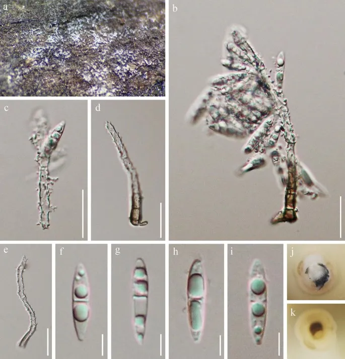
Pseudodactylaria camporesiana (MFLU 18-1535, holotype). a Colonies on submerged wood. b Conidiophore, conidiogenous cell and attached conidia. c – e Upper part of conidiophores and conidiogenous cells. f – i Conidia. j Colony on PDA (from front). k Colony on PDA (from reverse). Scale bars: b – e = 20 μm, f – i = 5 μm
Jenis
Pelat ilmiah
Kredit
Penulis dan sumber artikel: Hyde, Kevin D.; Dong, Yang; Phookamsak, Rungtiwa; Jeewon, Rajesh; Bhat, D. Jayarama; Jones, E. B. Gareth; Liu, Ning-Guo; Abeywickrama, Pranami D.; Mapook, Ausana; Wei, Deping; Perera, Rekhani H.; Manawasinghe, Ishara S.; Pem, Dhandevi; Bundhun, Digvijayini; Karunarathna, Anuruddha; Ekanayaka, Anusha H.; Bao, Dan-Feng; Li, Junfu; Samarakoon, Milan C.; Chaiwan, Napalai; Lin, Chuan-Gen; Phutthacharoen, Kunthida; Zhang, Sheng-Nan; Senanayake, Indunil C.; Goonasekara, Ishani D.; Thambugala, Kasun M.; Phukhamsakda, Chayanard; Tennakoon, Danushka S.; Jiang, Hong-Bo; Yang, Jing; Zeng, Ming; Huanraluek, Naruemon; Liu, Jian-Kui (Jack); Wijesinghe, Subodini N.; Tian, Qing; Tibpromma, Saowaluck; Brahmanage, Rashika S.; Boonmee, Saranyaphat; Huang, Shi-Ke; Thiyagaraja, Vinodhini; Lu, Yong-Zhong; Jayawardena, Ruvishika S.; Dong, Wei; Yang, Er-Fu; Singh, Sanjay K.; Singh, Shiv Mohan; Rana, Shiwali; Lad, Sneha S.; Anand, Garima; Devadatha, Bandarupalli; Niranjan, M.; Sarma, V. Venkateswara; Liimatainen, Kare; Aguirre-Hudson, Begoña; Niskanen, Tuula; Overall, Andy; Alvarenga, Renato Lúcio Mendes; Gibertoni, Tatiana Baptista; Pfliegler, Walter P.; Horváth, Enikő; Imre, Alexandra; Alves, Amanda Lucia; da Silva Santos, Ana Carla; Tiago, Patricia Vieira; Bulgakov, Timur S.; Wanasinghe, Dhanushaka N.; Bahkali, Ali H.; Doilom, Mingkwan; Elgorban, Abdallah M.; Maharachchikumbura, Sajeewa S. N.; Rajeshkumar, Kunhiraman C.; Haelewaters, Danny; Mortimer, Peter E.; Zhao, Qi; Lumyong, Saisamorn; Xu, Jianchu; Sheng, Jun
Saprobic on decaying, submerged wood in freshwater.
Sexual morph Undetermined.
Asexual morph Colonies on natural substrate, superficial, effuse, gregarious, white.
Mycelium partly immersed in natural substratum, composed of hyaline, branched, septate hyphae.
Conidiophores 35–45 × 3.5–5 µm (\(\bar{x} \) = 40 × 4.2 µm, n = 5), macronematous, mononematous, unbranched, subcylindrical, robust at base, straight to flexuous, rough, brown at the base, hyaline at upper part, septate, constricted at septa, thin-walled.
Conidiogenous cells 20–30 × 4–4.6 µm (\(\bar{x} \) = 26 × 4.2 µm, n = 5), holoblastic, polyblastic, integrated, terminal, sympodial, subcylindrical with apical taper, straight or flexuous, apical part forming a rachis with numerous, aggregated, cylindrical denticles, hyaline, rough-walled.
Conidia 18–22 × 3.5–4.5 µm (\(\bar{x} \) = 20 × 3.8 µm, n = 20), solitary, aggregating in slimy mass, pleuroacrogenous, narrowly fusiform, hyaline, smooth, guttulate, 1-septate, slightly constricted at the septa, lacking a sheath.
Culture characteristics: On PDA, colony circular, reaching 15 mm in 10 days at 25 °C, white from above, pale yellow from below, surface smooth with sparse mycelium, dry, edge entire, mycelium mostly immersed in culture.
Pseudodactylaria xanthorrhoeae and P. hyalotunicata have somewhat longer or thinner conidia in comparison with P. camporesiana (Tsui et al. 1997; Crous et al. 2017), and both species have a hyaline gelatinous sheath surrounding the conidia, a feature that is lacking in P. camporesiana.
Pseudodactylaria brevis is similar to P. camporesiana in having hyaline, 1-septate conidia without a sheath.
Saprobic on decaying, submerged wood in freshwater.
Sexual morph Undetermined.
Asexual morph Colonies on natural substrate, superficial, effuse, gregarious, white.
Mycelium partly immersed in natural substratum, composed of hyaline, branched, septate hyphae.
Conidiophores 35–45 × 3.5–5 µm (\(\bar{x} \) = 40 × 4.2 µm, n = 5), macronematous, mononematous, unbranched, subcylindrical, robust at base, straight to flexuous, rough, brown at the base, hyaline at upper part, septate, constricted at septa, thin-walled.
Conidiogenous cells 20–30 × 4–4.6 µm (\(\bar{x} \) = 26 × 4.2 µm, n = 5), holoblastic, polyblastic, integrated, terminal, sympodial, subcylindrical with apical taper, straight or flexuous, apical part forming a rachis with numerous, aggregated, cylindrical denticles, hyaline, rough-walled.
Conidia 18–22 × 3.5–4.5 µm (\(\bar{x} \) = 20 × 3.8 µm, n = 20), solitary, aggregating in slimy mass, pleuroacrogenous, narrowly fusiform, hyaline, smooth, guttulate, 1-septate, slightly constricted at the septa, lacking a sheath.
Culture characteristics: On PDA, colony circular, reaching 15 mm in 10 days at 25 °C, white from above, pale yellow from below, surface smooth with sparse mycelium, dry, edge entire, mycelium mostly immersed in culture.
Status: impor sumber; pemeriksaan editorial per profil belum selesai. Untuk klaim biologis, gunakan rujukan pada bagian terkait dan periksa konteks sumber aslinya.
Culture characteristics (14 days di 25 °C): Koloni pada PDA 15–16 mm dengan diameter, putih hingga pink pucat, tinggi, berduhai, tidak teratur, menghasilkan eksudat caesius yang banyak; kebalikan: cokelat hingga kayu manis. Koloni di MEA…
Koloni G. pannorum berwarna kuning-kecoklatan, dan biasanya memiliki tekstur granular, bubuk yang dihasilkan oleh struktur berspurulasi seperti pohon yang mikroskopis. The conidia of this fungus are small, wedge-shaped with a flat base. Th…
Spider host sepenuhnya ditutupi dengan mycelium putih. Conidiophores mononematous, muncul dari sisi hifa. Koloni tidak teratur pada PDA, 1.8–2.8 cm diam. setelah 14 d pada 25 °C, putih, terdiri dari felt basal, floccose hifa tumbuh…