01 / Morfologi

Ciri dan struktur

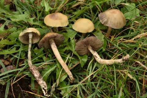
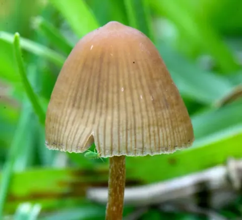
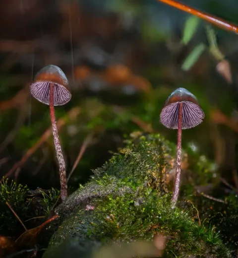
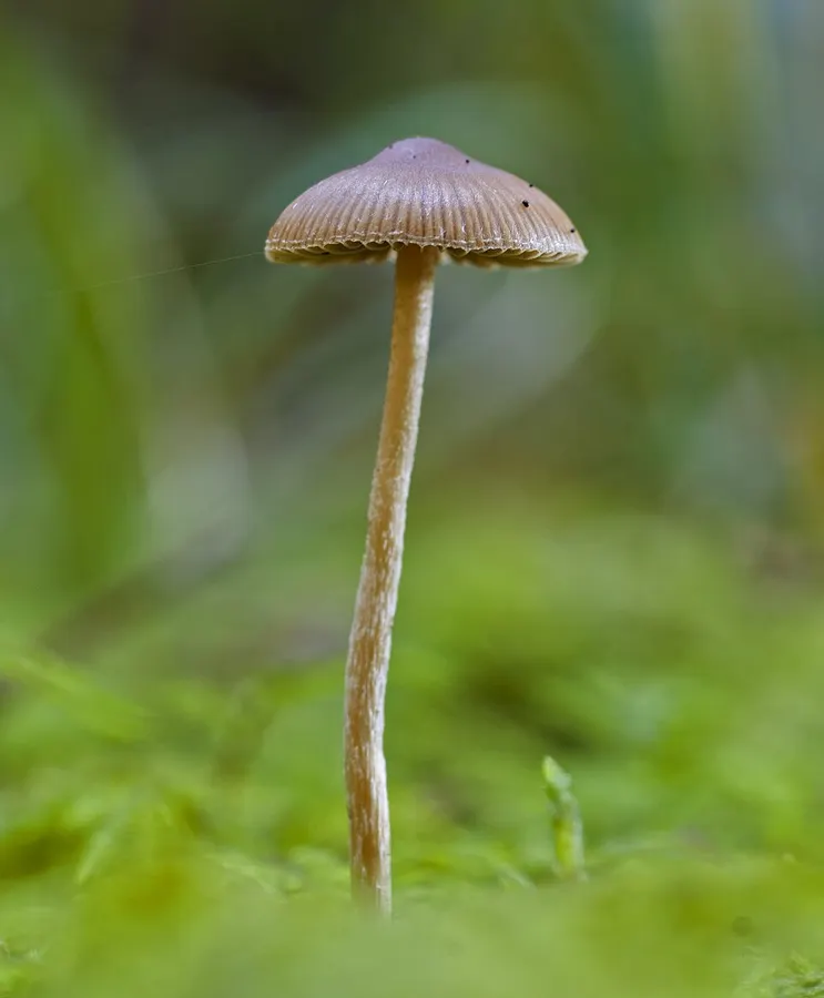
Psilocybe medullosa produces small, conical to bell-shaped caps (pilei) 1–2 cm across. The surface is slightly viscid but not peelable, with radial striations at the margin and an overall reddish-brown hue that pales slightly toward the edge. It is not strongly hygrophanous, although the colour may fade as it dries. A sparse, ephemeral (temporary) partial veil may be visible as fine fibrils on the cap margin of very young specimens. The gills (lamellae) are moderately crowded, with occasional short gills (lamellulae), ascending and narrowly attached (adnate) to the stipe; they are pale brown with a white edge. The slender stipe measures 5–8 cm by 0.2–0.3 cm, is hollow and slightly sinuous, and is pale brown with a paler tip. Its lower three-quarters are covered in conspicuous white fibrils, and the base often bears a tuft of fine mycelium. The flesh has a faint radish-like (raphanoid) odour. Microscopically, the spores are smooth, moderately thick-walled and measure 8–11 × 4.5–5.5 micrometres (μm). In face view they are ellipsoid-oblong, while in side view they appear flattened or somewhat almond-shaped, each bearing a small apical pore; they appear yellowish-brown in water and remain unchanged or only very faintly browned in Melzer's reagent. The basidia are four-spored. Cheilocystidia are numerous, 25–40 × 8–12 μm, flask-shaped with a usually narrow, elongated neck that may be straight or curved; pleurocystidia are absent. The cap cuticle (pileipellis) is an ixotrichoderm, consisting of a thin layer of loosely interwoven, gelatinised hyphae. Clamp connections occur throughout the hyphal system.

Sebagian uraian ditampilkan dalam bahasa sumber; terjemahannya belum tersedia atau belum lolos pemeriksaan.

Rujukan: [1]

Pengukuran mengikuti objek, spesimen atau kultur, dan metode yang disebutkan sumber. Baca uraian asli untuk konteks lengkap.

02 / Kehidupan

Habitat dan peran ekologis

Psilocybe medullosa is saprotrophic, fruiting in groups (gregariously) among decaying conifer litter—primarily needles and remnants of cones—in moss-rich coniferous woodland. The type collection was made on 14 September 2012 in a mossy spruce forest at Fiera di Primiero, Trentino, northern Italy. Although first described by Giacomo Bresadola in 1898, this species remains little reported in the Italian funga and has only sporadic published records from Lombardy and neighbouring Switzerland. The Trentino record thus represents one of the few confirmed Italian occurrences and helps to clarify its southern Alpine distribution.

Sebagian uraian ditampilkan dalam bahasa sumber; terjemahannya belum tersedia atau belum lolos pemeriksaan.

Rujukan: [1]

Habitat atau konteks temuan
hutan
Substrat
Informasi belum tersedia pada sumber rilis ini.
Peran atau konteks
Pengurai
Temukan hubungan serupa
03 / Siklus hidup

Reproduksi

Microscopically, the spores are smooth, moderately thick-walled and measure 8–11 × 4.5–5.5 micrometres (μm). In face view they are ellipsoid-oblong, while in side view they appear flattened or somewhat almond-shaped, each bearing a small apical pore; they appear yellowish-brown in water and remain unchanged or only very faintly browned in Melzer's reagent. The basidia are four-spored. Cheilocystidia are numerous, 25–40 × 8–12 μm, flask-shaped with a usually narrow, elongated neck that may be straight or curved; pleurocystidia are absent. The cap cuticle (pileipellis) is an ixotrichoderm, consisting of a thin layer of loosely interwoven, gelatinised hyphae. Clamp connections occur throughout the hyphal system.

Sebagian uraian ditampilkan dalam bahasa sumber; terjemahannya belum tersedia atau belum lolos pemeriksaan.

Rujukan: [1]

Sinonim menurut acuan taksonomi rilis: Galerina medullosa; Naucoria medullosa; Phaeogalera medullosa
04 / Batas informasi

Catatan identitas dan cakupan

  • Nama diterima mengikuti Catalogue of Life Base2026-09-11; uraian mempertahankan konsep dan nama sumber aslinya.
  • Original geographic, specimen and life-cycle scope retained; source import awaits individual editorial review.
  • Impor dan terjemahan otomatis belum diperiksa editorial per profil. Struktur reproduktif yang disebutkan tidak membuktikan seluruh siklus hidup diketahui.
  • Pengukuran pada uraian mengikuti spesimen, kultur, dan metode sumber; angka tidak diperluas ke semua populasi.

Catatan distribusi dalam narasi mengikuti sumbernya. Dokumentasi spesimen atau foto mengikuti catatan sumber. Bukti wilayah Indonesia dan penilaian konservasi tidak disimpulkan dari gambar.

05 / Dokumentasi

Melihat dengan konteks

Dokumentasi Psilocybe medullosa dari Wikimedia Commons. For more information about this, see the observation page at Mushroom Observer. Identifikasi mengikuti record berkas sumber.

Jenis
Fotografi
Kredit
This image was created by user Gerhard_S at Mushroom Observer , a source for mycological images. You can contact this user here .
Sumber media
Buka entri sumber

Gambar menunjukkan objek menurut identifikasi sumber, bukan verifikasi independen spesimen oleh Fungi. Tidak ada skala baru yang ditambahkan.

06 / Rujukan

Sumber profil

  1. [1] Psilocybe medullosa — Wikipedia (en)Bagian sumber: Frozen revision 1364422986
    Tanggal penghimpunan sumber: 2026-10-12
    Teks sumber: CC BY-SA4.0 · Wikipedia contributors
  2. [2] Catalogue of Life Base2026-09-11 — Psilocybe medullosaBagian sumber: ColDP NameUsage.tsv ID 4PH8K; accepted species; source 2073
    Tanggal penghimpunan sumber: 2026-10-12
  3. [3] Dokumentasi visual — Psilocybe medullosaBagian sumber: Individual media record; identification follows the source, not independent determination
    Tanggal penghimpunan sumber: 2026-10-12
    Media sumber: CC BY-SA 3.0 · This image was created by user Gerhard_S at Mushroom Observer , a source for mycological images. You can contact this user here .

Adaptasi teks: CC BY-SA 4.0. Wikipedia contributors — Psilocybe medullosa — Wikipedia (en)

Uraian asli dan riwayat sumber

Riwayat atau halaman sumber

Ringkasan sumber

Psilocybe medullosa is a species of psychoactive mushroom. A widespread but rather rare species, it is found in Europe, where it grows as a saprobe on woody debris and detritus. Chemical analysis has been used to confirm the presence of the psychedelic compounds psilocin and psilocybin in the fruit bodies but probably at low levels.

Revisi sumber

Morfologi sumber

Psilocybe medullosa produces small, conical to bell-shaped caps (pilei) 1–2 cm across. The surface is slightly viscid but not peelable, with radial striations at the margin and an overall reddish-brown hue that pales slightly toward the edge. It is not strongly hygrophanous, although the colour may fade as it dries. A sparse, ephemeral (temporary) partial veil may be visible as fine fibrils on the cap margin of very young specimens. The gills (lamellae) are moderately crowded, with occasional short gills (lamellulae), ascending and narrowly attached (adnate) to the stipe; they are pale brown with a white edge. The slender stipe measures 5–8 cm by 0.2–0.3 cm, is hollow and slightly sinuous, and is pale brown with a paler tip. Its lower three-quarters are covered in conspicuous white fibrils, and the base often bears a tuft of fine mycelium. The flesh has a faint radish-like (raphanoid) odour. Microscopically, the spores are smooth, moderately thick-walled and measure 8–11 × 4.5–5.5 micrometres (μm). In face view they are ellipsoid-oblong, while in side view they appear flattened or somewhat almond-shaped, each bearing a small apical pore; they appear yellowish-brown in water and remain unchanged or only very faintly browned in Melzer's reagent. The basidia are four-spored. Cheilocystidia are numerous, 25–40 × 8–12 μm, flask-shaped with a usually narrow, elongated neck that may be straight or curved; pleurocystidia are absent. The cap cuticle (pileipellis) is an ixotrichoderm, consisting of a thin layer of loosely interwoven, gelatinised hyphae. Clamp connections occur throughout the hyphal system.

Revisi sumber

Ekologi sumber

Psilocybe medullosa is saprotrophic, fruiting in groups (gregariously) among decaying conifer litter—primarily needles and remnants of cones—in moss-rich coniferous woodland. The type collection was made on 14 September 2012 in a mossy spruce forest at Fiera di Primiero, Trentino, northern Italy. Although first described by Giacomo Bresadola in 1898, this species remains little reported in the Italian funga and has only sporadic published records from Lombardy and neighbouring Switzerland. The Trentino record thus represents one of the few confirmed Italian occurrences and helps to clarify its southern Alpine distribution.

Revisi sumber

Struktur reproduksi sumber

Microscopically, the spores are smooth, moderately thick-walled and measure 8–11 × 4.5–5.5 micrometres (μm). In face view they are ellipsoid-oblong, while in side view they appear flattened or somewhat almond-shaped, each bearing a small apical pore; they appear yellowish-brown in water and remain unchanged or only very faintly browned in Melzer's reagent. The basidia are four-spored. Cheilocystidia are numerous, 25–40 × 8–12 μm, flask-shaped with a usually narrow, elongated neck that may be straight or curved; pleurocystidia are absent. The cap cuticle (pileipellis) is an ixotrichoderm, consisting of a thin layer of loosely interwoven, gelatinised hyphae. Clamp connections occur throughout the hyphal system.

Revisi sumber

Rujukan halaman ini

Fungi. Psilocybe medullosa. Rilis data fungi-2026-10-12-expansion-2. https://fungi-atlas.mgadib.chatgpt.site/jamur/psilocybe-medullosa/.

Status: impor sumber; pemeriksaan editorial per profil belum selesai. Untuk klaim biologis, gunakan rujukan pada bagian terkait dan periksa konteks sumber aslinya.