Ciri dan struktur
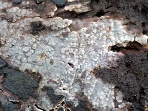
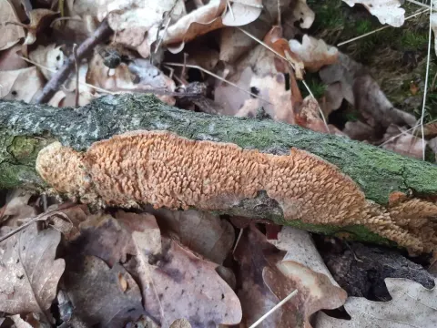
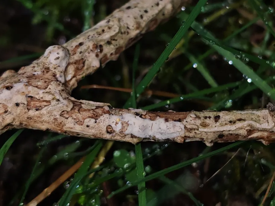
Hymenophore annual, ceraceous, adherent, effused forming linear areas to 20 x 4 cm; hymenial surface cream, becoming pinkish-buff, even, at length creviced; margin thinning out, somewhat pelliculose, cream, adherent. Context white, 100-500 µm thick, basal layer thick, of parallel compact hyphae, intermediate layer of erect hyphae compactly arranged and often cemented; generative hyphae 3-4 µm diameter, walls 0.2 µm thick, naked, with clamp connections. Hymenial layer to 40 µm deep, a dense palisade of basidia, paraphyses, and scanty paraphysate hyphae. Basidia subclavate, 25-35 x 7-9 µm, bearing 4 spores; sterigmata slender, to 6 µm long. Paraphyses.subclavate, 14-22 x 6-7 µm. Paraphysate hyphae cylindrical, to 4 µm diameter, projecting to 20 µm. Spores globose or subglobose, apiculate, 6-8 µm diameter, walls smooth, hyaline, 0.5 µm thick.
Habitat dan peran ekologis
DISTRIBUTION: Western Europe, Great Britain, Australia, New Zealand. HABITAT: Effused on bark of dead branches.
- Habitat atau konteks temuan
- Informasi belum tersedia pada sumber rilis ini.
- Substrat
- kulit kayu
- Peran atau konteks
- Belum dikategorikan pada rilis ini.
Reproduksi
Hymenophore annual, ceraceous, adherent, effused forming linear areas to 20 x 4 cm; hymenial surface cream, becoming pinkish-buff, even, at length creviced; margin thinning out, somewhat pelliculose, cream, adherent. Context white, 100-500 µm thick, basal layer thick, of parallel compact hyphae, intermediate layer of erect hyphae compactly arranged and often cemented; generative hyphae 3-4 µm diameter, walls 0.2 µm thick, naked, with clamp connections. Hymenial layer to 40 µm deep, a dense palisade of basidia, paraphyses, and scanty paraphysate hyphae. Basidia subclavate, 25-35 x 7-9 µm, bearing 4 spores; sterigmata slender, to 6 µm long. Paraphyses.subclavate, 14-22 x 6-7 µm. Paraphysate hyphae cylindrical, to 4 µm diameter, projecting to 20 µm.
Catatan identitas dan cakupan
- Nama diterima mengikuti Catalogue of Life Base2026-09-11; uraian mempertahankan konsep dan nama sumber aslinya.
- Historical description as cited by BiotaNZ. Preserve explicit New Zealand/locality, host and specimen qualifiers. Distribution is the cited source scope, not a current global range.
- Impor dan terjemahan otomatis belum diperiksa editorial per profil. Struktur reproduktif yang disebutkan tidak membuktikan seluruh siklus hidup diketahui.
- Pengukuran pada uraian mengikuti spesimen, kultur, dan metode sumber; angka tidak diperluas ke semua populasi.
Catatan distribusi dalam narasi mengikuti sumbernya. Dokumentasi spesimen atau foto mengikuti catatan sumber. Bukti wilayah Indonesia dan penilaian konservasi tidak disimpulkan dari gambar.
Melihat dengan konteks
Dokumentasi Radulomyces rickii menurut asosiasi foto pada record takson iNaturalist. Kredit: Bohan Jia. Identitas mengikuti sumber; bukan penentuan spesimen independen.
- Jenis
- Fotografi
- Kredit
- (c) Bohan Jia, some rights reserved (CC BY)
- Lisensi
- CC BY 4.0
- Sumber media
- Buka entri sumber
Dokumentasi tambahan
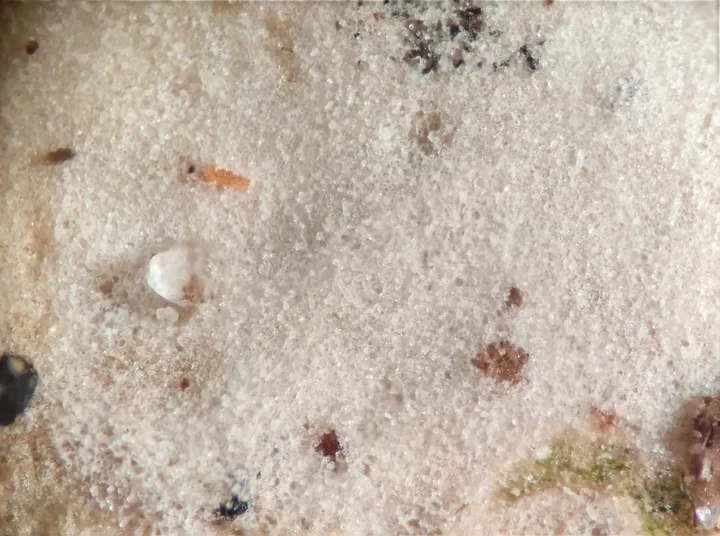
Foto yang diasosiasikan dengan Radulomyces rickii pada iNaturalist. Identifikasi mengikuti sumber; bukan penentuan spesimen independen.
Kredit dan penggunaan
(c) Bohan Jia, some rights reserved (CC BY)
Ukuran dan format gambar disesuaikan tanpa pemotongan, retus, atau penambahan skala. Lisensi sumber dipertahankan.
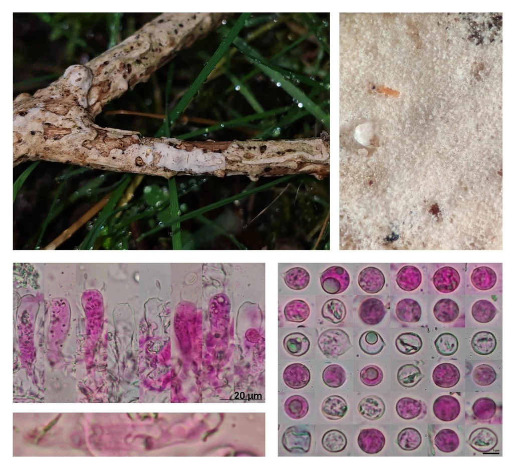
Pelat foto makro material jamur dan mikrograf yang diasosiasikan dengan Radulomyces rickii pada iNaturalist. Identitas mengikuti sumber.
Kredit dan penggunaan
(c) Bohan Jia, some rights reserved (CC BY)
Ukuran dan format gambar disesuaikan tanpa pemotongan, retus, atau penambahan skala. Lisensi sumber dipertahankan.
Sumber profil
- [1] Cunningham, G.H. 1963: The Thelephoraceae of Australia and New Zealand. New Zealand Department of Scientific and Industrial Research, Bulletin. 145. — Corticium rickii
- [2] Catalogue of Life Base2026-09-11 — Radulomyces rickii
- [3] Dokumentasi visual — Radulomyces rickii
- [4] Dokumentasi visual — Radulomyces rickii
- [5] Dokumentasi visual — Radulomyces rickii
Rujukan halaman ini
Fungi. Radulomyces rickii. Rilis data fungi-2026-10-12-expansion-2. https://fungi-atlas.mgadib.chatgpt.site/jamur/radulomyces-rickii/.
Status: impor sumber; pemeriksaan editorial per profil belum selesai. Untuk klaim biologis, gunakan rujukan pada bagian terkait dan periksa konteks sumber aslinya.