Ciri dan struktur
Saprobic on a dead leaf of Musa sp. Sexual morph Undetermined. Asexual morph Hyphomycetous. Sporodochia dark, dense, dry, powdery, velvety, 1–2 mm diameter. Conidiophore mother cells 3.4–5.8 × 3.7–4.7 μm (x̄ = 4.6 × 4.1 μm, n = 10) subhyaline or light brown, doliiform or subspherical. Conidiophores usually short to long bearing two types of conidia referred to here as α and β. Conidiophores of α conidia up to 40–85 × 0.8–2.5 μm (x̄ = 64 × 21.7 μm, n = 15), pale brown or dark golden brown, rough-walled, hyaline at bottom near the conidiophore mother cell, pale brown at middle, dark golden brown at top near conidial cells, erect or flexuous, narrow and long, generally unbranched, rarely branched. Conidiophores of β conidia 0.7–3.5 × 1.5–3 μm (x̄ = 1.9 × 2.3 μm, n = 15) short, erect, unbranched, hyaline when immature, subhyaline or hyaline at maturity. Conidiogenous cell development basauxic, forming a single, terminal holoblastic conidium at the apex of conidiophore. Conidial development holoblastic. Conidia solitary, dry, two types: α conidia stellate, 15–22.7 × 14.5–20.5 μm (x̄ = 18.8 × 17.8 μm, n = 15), 4–6 celled, each cell globose to subglobose, deeply constricted at the septa, conspicuously spinulate, 4–6 spines, each 2–8 μm long arise from surface of each cell. β conidia disc-shaped, initially hyaline, 4-celled, each cell slightly turbinate in shape, rough-walled, crossed septate, becoming brown to dark brown at maturity, each cell turbinate, crossed-septate, smooth-walled, light brown at the center near the septa, dark brown at periphery in constricted areas, 9.3–14.2 × 8.4–12.5 μm (x̄ = 12.7 × 10.8 μm, n = 40), somewhat obovoid, deeply constricted at the septa, flat from side view, frequently with attached conidiogenous cells when splitting from the conidiophores.
Habitat dan peran ekologis
Saprobic on a dead leaf of Musa sp. Sexual morph Undetermined. Asexual morph Hyphomycetous. Sporodochia dark, dense, dry, powdery, velvety, 1–2 mm diameter. Conidiophore mother cells 3.4–5.8 × 3.7–4.7 μm (x̄ = 4.6 × 4.1 μm, n = 10) subhyaline or light brown, doliiform or subspherical. Conidiophores usually short to long bearing two types of conidia referred to here as α and β. Conidiophores of α conidia up to 40–85 × 0.8–2.5 μm (x̄ = 64 × 21.7 μm, n = 15), pale brown or dark golden brown, rough-walled, hyaline at bottom near the conidiophore mother cell, pale brown at middle, dark golden brown at top near conidial cells, erect or flexuous, narrow and long, generally unbranched, rarely branched. Conidiophores of β conidia 0.7–3.5 × 1.5–3 μm (x̄ = 1.9 × 2.3 μm, n = 15) short, erect, unbranched, hyaline when immature, subhyaline or hyaline at maturity. Conidiogenous cell development basauxic, forming a single, terminal holoblastic conidium at the apex of conidiophore. Conidial development holoblastic. Conidia solitary, dry, two types: α conidia stellate, 15–22.7 × 14.5–20.5 μm (x̄ = 18.8 × 17.8 μm, n = 15), 4–6 celled, each cell globose to subglobose, deeply constricted at the septa, conspicuously spinulate, 4–6 spines, each 2–8 μm long arise from surface of each cell. β conidia disc-shaped, initially hyaline, 4-celled, each cell slightly turbinate in shape, rough-walled, crossed septate, becoming brown to dark brown at maturity, each cell turbinate, crossed-septate, smooth-walled, light brown at the center near the septa, dark brown at periphery in constricted areas, 9.3–14.2 × 8.4–12.5 μm (x̄ = 12.7 × 10.8 μm, n = 40), somewhat obovoid, deeply constricted at the septa, flat from side view, frequently with attached conidiogenous cells when splitting from the conidiophores.
- Habitat atau konteks temuan
- Informasi belum tersedia pada sumber rilis ini.
- Substrat
- Informasi belum tersedia pada sumber rilis ini.
- Peran atau konteks
- Pengurai
Reproduksi
Saprobic on a dead leaf of Musa sp. Sexual morph Undetermined. Asexual morph Hyphomycetous. Sporodochia dark, dense, dry, powdery, velvety, 1–2 mm diameter. Conidiophore mother cells 3.4–5.8 × 3.7–4.7 μm (x̄ = 4.6 × 4.1 μm, n = 10) subhyaline or light brown, doliiform or subspherical. Conidiophores usually short to long bearing two types of conidia referred to here as α and β. Conidiophores of α conidia up to 40–85 × 0.8–2.5 μm (x̄ = 64 × 21.7 μm, n = 15), pale brown or dark golden brown, rough-walled, hyaline at bottom near the conidiophore mother cell, pale brown at middle, dark golden brown at top near conidial cells, erect or flexuous, narrow and long, generally unbranched, rarely branched.
Catatan identitas dan cakupan
- Nama diterima mengikuti Catalogue of Life Base2026-09-11; uraian mempertahankan konsep dan nama sumber aslinya.
- Preserve original treatment localities, hosts and substrate scope. Materials examined describe documented specimens, not the full global range.
- Impor dan terjemahan otomatis belum diperiksa editorial per profil. Struktur reproduktif yang disebutkan tidak membuktikan seluruh siklus hidup diketahui.
- Pengukuran pada uraian mengikuti spesimen, kultur, dan metode sumber; angka tidak diperluas ke semua populasi.
Catatan distribusi dalam narasi mengikuti sumbernya. Dokumentasi spesimen atau foto mengikuti catatan sumber. Bukti wilayah Indonesia dan penilaian konservasi tidak disimpulkan dari gambar.
Melihat dengan konteks
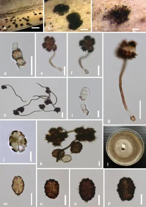
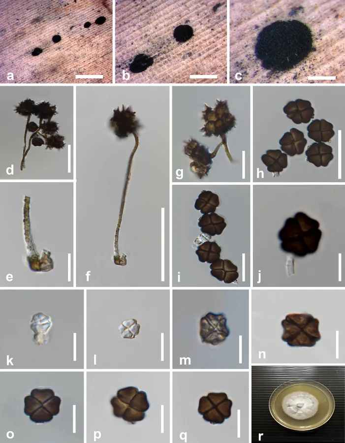
Spegazzinia musae (MFLU 19-2907, holotype) a–c fungal colonies on host surface d mature conidia e conidiophore of α conidia with the mother cell f, g α conidia h–q β conidia r colony on PDA after 28 days. Scale bars: 200 μm (a–c), 20 μm (d–g, j), 10 μm (h, i, k–q).
- Jenis
- Pelat ilmiah
- Kredit
- Binu C. Samarakoon, Rungtiwa Phookamsak, Dhanushka N. Wanasinghe, Putarak Chomnunti, Kevin D. Hyde, Eric H. C. Mckenzie, Itthayakorn Promputtha, Jian-Chu Xu, Yun-Ju Li
- Lisensi
- CC BY 4.0
- Sumber media
- Buka entri sumber
Sumber profil
- [1] Taxonomy and phylogenetic appraisal of Spegazzinia musae sp. nov. and S. deightonii (Didymosphaeriaceae, Pleosporales) on Musaceae from Thailand — Spegazzinia musae
- [2] Catalogue of Life Base2026-09-11 — Spegazzinia musae
- [3] Dokumentasi visual — Spegazzinia musae
Rujukan halaman ini
Fungi. Spegazzinia musae. Rilis data fungi-2026-10-12-expansion-2. https://fungi-atlas.mgadib.chatgpt.site/jamur/spegazzinia-musae/.
Status: impor sumber; pemeriksaan editorial per profil belum selesai. Untuk klaim biologis, gunakan rujukan pada bagian terkait dan periksa konteks sumber aslinya.