Ciri dan struktur
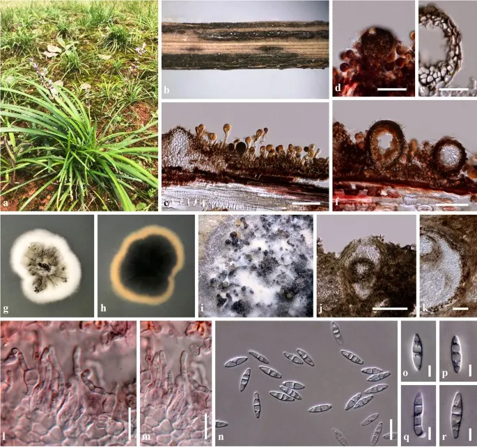
Associated with rust on living leaves of Liriope spicata (Thunb.) Lour. Sexual morph Undetermined. Asexual morph Coelomycetous. Conidiomata 105–160 µm high, 90–150 µm diam., black, pycnidial, solitary, associated with rust stromatic along the leaf veins, semi-immersed to superficial on stromata, globose to subglobose, uni-loculate, glabrous, ostiole central, with pore-like opening. Conidiomata walls 12–30 µm wide, composed of 2–5 layers, of dark brown pseudoparenchymatous cells of textura angularis. Conidiophores 15–21 × 2.4–3 µm (\(\bar{x} \) = 18 × 2.7 µm, n = 20), arising from the basal cavity, 1–2-celled, hyaline, curved, cylindrical, or reduced to conidiogenous cells. Conidiogenou s cells 3.5–6 × 2.5–4.5 µm (\(\bar{x} \) = 4.7 × 3.5 µm, n = 20), enteroblastic, phialidic, discrete, determinate, cylindrical to ampulliform to doliiform, hyaline, 0–1-septate, smooth, thin-walled, minute collarette, with 1–2 apertures, and periclinal wall thickening. Conidia 14–17 × 3–5 µm (\(\bar{x} \) = 15.5 × 4.5 µm, n = 20), hyaline, fusiform to ellipsoidal, mostly 1–3-septate, constricted at the central septum, smooth-walled. Culture characteristics: Colonies on PDA reaching 25–30 mm diam. after 4 weeks at room temperature. Colony dense, irregular in shape, flattened, slightly raised, surface slightly rough, heaped and folded at the centre, with small granular and black, stromatic, edge undulate, with margin well-defined, felted at the centre, fluffy at the edge; from above white at the margin, with yellowish grey to greenish grey at the centre; from below, white to cream at the margin, black at the centre; not producing pigmentation on agar medium, sporulating on PDA after 3 weeks.
Habitat dan peran ekologis
Known hosts and distribution: Associated with rust on Pennisetum sp. (Brazil), on Ravenelia macowania on Vachellia karroo (South Africa), on leaves of Phragmites sp. (Australia), and associated with rust on living leaves of Liriope spicata (Yunnan, China) (Trakunyingcharoen et al. 2014; Crous et al. 2018; this study).
- Habitat atau konteks temuan
- Informasi belum tersedia pada sumber rilis ini.
- Substrat
- Informasi belum tersedia pada sumber rilis ini.
- Peran atau konteks
- Belum dikategorikan pada rilis ini.
Reproduksi
Associated with rust on living leaves of Liriope spicata (Thunb.) Lour. Sexual morph Undetermined. Asexual morph Coelomycetous. Conidiomata 105–160 µm high, 90–150 µm diam., black, pycnidial, solitary, associated with rust stromatic along the leaf veins, semi-immersed to superficial on stromata, globose to subglobose, uni-loculate, glabrous, ostiole central, with pore-like opening. Conidiomata walls 12–30 µm wide, composed of 2–5 layers, of dark brown pseudoparenchymatous cells of textura angularis. Conidiophores 15–21 × 2.4–3 µm (\(\bar{x} \) = 18 × 2.7 µm, n = 20), arising from the basal cavity, 1–2-celled, hyaline, curved, cylindrical, or reduced to conidiogenous cells. Conidiogenou s cells 3.5–6 × 2.5–4.5 µm (\(\bar{x} \) = 4.7 × 3.5 µm, n = 20), enteroblastic, phialidic, discrete, determinate, cylindrical to ampulliform to doliiform, hyaline, 0–1-septate, smooth, thin-walled, minute collarette, with 1–2 apertures, and periclinal wall thickening.
Catatan identitas dan cakupan
- Nama diterima mengikuti Catalogue of Life Base2026-09-11; uraian mempertahankan konsep dan nama sumber aslinya.
- Preserve original monograph treatment localities, hosts and specimen/substrate scope. Materials examined document specimens, not the full global range.
- Impor dan terjemahan otomatis belum diperiksa editorial per profil. Struktur reproduktif yang disebutkan tidak membuktikan seluruh siklus hidup diketahui.
- Pengukuran pada uraian mengikuti spesimen, kultur, dan metode sumber; angka tidak diperluas ke semua populasi.
Catatan distribusi dalam narasi mengikuti sumbernya. Dokumentasi spesimen atau foto mengikuti catatan sumber. Bukti wilayah Indonesia dan penilaian konservasi tidak disimpulkan dari gambar.
Melihat dengan konteks
Sphaerellopsis paraphysata (KUN-HKAS 101483). a Liriope spicata. b – d Appearance of conidiomata associated with rust on host substrate. e Section through conidioma wall. f Section through conidioma. g, h Culture on PDA after 2 weeks (g = from above, h = from below). i–r in vitro (OA). i Sporulation on OA after 4 weeks. j Section through conidioma. k Section through conidioma wall. l, m Conidiogenous cells stained in congo red. n–r Conidia. Scale bars j = 100 µm, c, d, f = 50 µm, k = 20 µm, e, l, m = 10 µm, n = 5 µm, o–r = 2 µm
- Jenis
- Pelat ilmiah
- Kredit
- Penulis dan sumber artikel: Phookamsak, Rungtiwa; Hyde, Kevin D.; Jeewon, Rajesh; Bhat, D. Jayarama; Jones, E. B. Gareth; Maharachchikumbura, Sajeewa S. N.; Raspé, Olivier; Karunarathna, Samantha C.; Wanasinghe, Dhanushka N.; Hongsanan, Sinang; Doilom, Mingkwan; Tennakoon, Danushka S.; Machado, Alexandre R.; Firmino, André L.; Ghosh, Aniket; Karunarathna, Anuruddha; Mešić, Armin; Dutta, Arun Kumar; Thongbai, Benjarong; Devadatha, Bandarupalli; Norphanphoun, Chada; Senwanna, Chanokned; Wei, Deping; Pem, Dhandevi; Ackah, Frank Kwekucher; Wang, Gen-Nuo; Jiang, Hong-Bo; Madrid, Hugo; Lee, Hyang Burm; Goonasekara, Ishani D.; Manawasinghe, Ishara S.; Kušan, Ivana; Cano, Josep; Gené, Josepa; Li, Junfu; Das, Kanad; Acharya, Krishnendu; Raj, K. N. Anil; Latha, K. P. Deepna; Chethana, K. W. Thilini; He, Mao-Qiang; Dueñas, Margarita; Jadan, Margita; Martín, María P.; Samarakoon, Milan C.; Dayarathne, Monika C.; Raza, Mubashar; Park, Myung Soo; Telleria, M. Teresa; Chaiwan, Napalai; Matočec, Neven; de Silva, Nimali I.; Pereira, Olinto L.; Singh, Paras Nath; Manimohan, Patinjareveettil; Uniyal, Priyanka; Shang, Qiu-Ju; Bhatt, Rajendra P.; Perera, Rekhani H.; Alvarenga, Renato Lúcio Mendes; Nogal-Prata, Sandra; Singh, Sanjay K.; Vadthanarat, Santhiti; Oh, Seung-Yoon; Huang, Shi-Ke; Rana, Shiwali; Konta, Sirinapa; Paloi, Soumitra; Jayasiri, Subashini C.; Jeon, Sun Jeong; Mehmood, Tahir; Gibertoni, Tatiana Baptista; Nguyen, Thuong T. T.; Singh, Upendra; Thiyagaraja, Vinodhini; Sarma, V. Venkateswara; Dong, Wei; Yu, Xian-Dong; Lu, Yong-Zhong; Lim, Young Woon; Chen, Yun; Tkalčec, Zdenko; Zhang, Zhi-Feng; Luo, Zong-Long; Daranagama, Dinushani A.; Thambugala, Kasun M.; Tibpromma, Saowaluck; Camporesi, Erio; Bulgakov, Timur S.; Dissanayake, Asha J.; Senanayake, Indunil C.; Dai, Dong Qin; Tang, Li-Zhou; Khan, Sehroon; Zhang, Huang; Promputtha, Itthayakorn; Cai, Lei; Chomnunti, Putarak; Zhao, Rui-Lin; Lumyong, Saisamorn; Boonmee, Saranyaphat; Wen, Ting-Chi; Mortimer, Peter E.; Xu, Jianchu
- Lisensi
- CC BY 4.0
- Sumber media
- Buka entri sumber
Sumber profil
- [1] Fungal diversity notes 929–1035: taxonomic and phylogenetic contributions on genera and species of fungi — Sphaerellopsis paraphysata
- [2] Catalogue of Life Base2026-09-11 — Sphaerellopsis paraphysata
- [3] Dokumentasi visual — Sphaerellopsis paraphysata
Rujukan halaman ini
Fungi. Sphaerellopsis paraphysata. Rilis data fungi-2026-10-12-expansion-2. https://fungi-atlas.mgadib.chatgpt.site/jamur/sphaerellopsis-paraphysata/.
Status: impor sumber; pemeriksaan editorial per profil belum selesai. Untuk klaim biologis, gunakan rujukan pada bagian terkait dan periksa konteks sumber aslinya.