01 / Morfologi

Ciri dan struktur

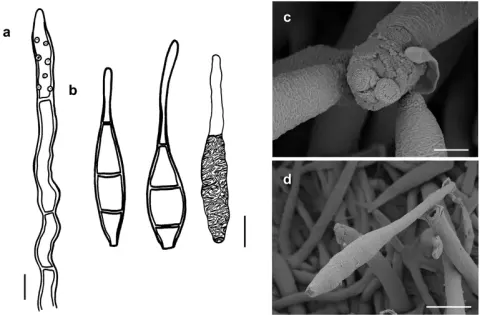
Colonies effuse, olive to olivaceous-brown, velvety or hairy. Hyphae superficial, branched, septate, 0.5–2 µm wide, pale olive to olivaceous-brown, smooth. Conidiophores arising singly or in groups terminally or laterally from the hyphae, erect or ascending, straight or flexous, septate, up to 400 µm long, 2–4 µm thick along most of their length, swollen towards the apex, 5–8 µm thick, brown, reticulate as seen by SEM, with scattered cylindrical scars. Conidia solitary, straight or slightly curved, obovate to clavate, truncate at their base, 3–septate, (10–)12–14(–15) × (5–)6–8 µm, 1.5–2 µm wide at the base, the cells at each end of a conidium subhyaline or pale brown, intermediate cells brown, ornamented. As seen by SEM, the ornamentation of the spores is distinctly reticulated, with thin to thick networks that can form ridges.

Sebagian uraian ditampilkan dalam bahasa sumber; terjemahannya belum tersedia atau belum lolos pemeriksaan.

Rujukan: [1]

Pengukuran mengikuti objek, spesimen atau kultur, dan metode yang disebutkan sumber. Baca uraian asli untuk konteks lengkap.

02 / Kehidupan

Habitat dan peran ekologis

On colonies of Meliola borneensis on living leaves of Uvaria chamae (Annonaceae) in Sierra Leone (Ellis 1968).

Sebagian uraian ditampilkan dalam bahasa sumber; terjemahannya belum tersedia atau belum lolos pemeriksaan.

Rujukan: [1]

Habitat atau konteks temuan
Informasi belum tersedia pada sumber rilis ini.
Substrat
Informasi belum tersedia pada sumber rilis ini.
Peran atau konteks
Belum dikategorikan pada rilis ini.
Temukan hubungan serupa
03 / Siklus hidup

Reproduksi

Colonies effuse, olive to olivaceous-brown, velvety or hairy. Hyphae superficial, branched, septate, 0.5–2 µm wide, pale olive to olivaceous-brown, smooth. Conidiophores arising singly or in groups terminally or laterally from the hyphae, erect or ascending, straight or flexous, septate, up to 400 µm long, 2–4 µm thick along most of their length, swollen towards the apex, 5–8 µm thick, brown, reticulate as seen by SEM, with scattered cylindrical scars. Conidia solitary, straight or slightly curved, obovate to clavate, truncate at their base, 3–septate, (10–)12–14(–15) × (5–)6–8 µm, 1.5–2 µm wide at the base, the cells at each end of a conidium subhyaline or pale brown, intermediate cells brown, ornamented.

Sebagian uraian ditampilkan dalam bahasa sumber; terjemahannya belum tersedia atau belum lolos pemeriksaan.

Rujukan: [1]

04 / Batas informasi

Catatan identitas dan cakupan

  • Nama diterima mengikuti Catalogue of Life Base2026-09-11; uraian mempertahankan konsep dan nama sumber aslinya.
  • Preserve original treatment localities, hosts and substrate scope. Materials examined describe documented specimens, not the full global range.
  • Impor dan terjemahan otomatis belum diperiksa editorial per profil. Struktur reproduktif yang disebutkan tidak membuktikan seluruh siklus hidup diketahui.
  • Pengukuran pada uraian mengikuti spesimen, kultur, dan metode sumber; angka tidak diperluas ke semua populasi.

Catatan distribusi dalam narasi mengikuti sumbernya. Dokumentasi spesimen atau foto mengikuti catatan sumber. Bukti wilayah Indonesia dan penilaian konservasi tidak disimpulkan dari gambar.

05 / Dokumentasi

Melihat dengan konteks

Spiropes deightonii (IMI48956a) a conidiophores b conidia, as seen by LM (two upper spores; the thickness of the wall is indicated only in the drawing on the left-hand side) and by SEM (bottom spore) c, d as seen by SEM c conidiophore d conidia. Scale bars: 5 μm (a, b); 8 μm (c); 5 μm (d).

Jenis
Pelat ilmiah
Kredit
Miguel A. Bermúdez-Cova, Tina A. Hofmann, Nourou S. Yorou, Meike Piepenbring
Lisensi
CC BY 4.0
Sumber media
Buka entri sumber

Gambar menunjukkan objek menurut identifikasi sumber, bukan verifikasi independen spesimen oleh Fungi. Tidak ada skala baru yang ditambahkan.

06 / Rujukan

Sumber profil

  1. [1] Systematic revision of species of Atractilina and Spiropes hyperparasitic on Meliolales (Ascomycota) in the tropics — Spiropes deightoniiBagian sumber: PMC11031638_section_67
    Tanggal penghimpunan sumber: 2026-10-12
    Teks sumber: CC BY 4.0 · Bermúdez-Cova MA, Hofmann TA, Yorou NS, Piepenbring M.
  2. [2] Catalogue of Life Base2026-09-11 — Spiropes deightoniiBagian sumber: ColDP NameUsage.tsv ID 4Z897; accepted species; source 2073
    Tanggal penghimpunan sumber: 2026-10-12
  3. [3] Dokumentasi visual — Spiropes deightoniiBagian sumber: Individual media record; identification follows the source, not independent determination
    Tanggal penghimpunan sumber: 2026-10-12
    Media sumber: CC BY 4.0 · Miguel A. Bermúdez-Cova, Tina A. Hofmann, Nourou S. Yorou, Meike Piepenbring

Adaptasi teks: CC BY 4.0. Bermúdez-Cova MA, Hofmann TA, Yorou NS, Piepenbring M. — Systematic revision of species of Atractilina and Spiropes hyperparasitic on Meliolales (Ascomycota) in the tropics — Spiropes deightonii

Uraian asli dan riwayat sumber

Riwayat atau halaman sumber

Ringkasan sumber

Colonies effuse, olive to olivaceous-brown, velvety or hairy. Hyphae superficial, branched, septate, 0.5–2 µm wide, pale olive to olivaceous-brown, smooth. Conidiophores arising singly or in groups terminally or laterally from the hyphae, erect or ascending, straight or flexous, septate, up to 400 µm long, 2–4 µm thick along most of their length, swollen towards the apex, 5–8 µm thick, brown, reticulate as seen by SEM, with scattered cylindrical scars. Conidia solitary, straight or slightly curved, obovate to clavate, truncate at their base, 3–septate, (10–)12–14(–15) × (5–)6–8 µm, 1.5–2 µm wide at the base, the cells at each end of a conidium subhyaline or pale brown, intermediate cells brown, ornamented.

Revisi sumber

Morfologi sumber

Colonies effuse, olive to olivaceous-brown, velvety or hairy. Hyphae superficial, branched, septate, 0.5–2 µm wide, pale olive to olivaceous-brown, smooth. Conidiophores arising singly or in groups terminally or laterally from the hyphae, erect or ascending, straight or flexous, septate, up to 400 µm long, 2–4 µm thick along most of their length, swollen towards the apex, 5–8 µm thick, brown, reticulate as seen by SEM, with scattered cylindrical scars. Conidia solitary, straight or slightly curved, obovate to clavate, truncate at their base, 3–septate, (10–)12–14(–15) × (5–)6–8 µm, 1.5–2 µm wide at the base, the cells at each end of a conidium subhyaline or pale brown, intermediate cells brown, ornamented. As seen by SEM, the ornamentation of the spores is distinctly reticulated, with thin to thick networks that can form ridges.

Revisi sumber

Ekologi sumber

On colonies of Meliola borneensis on living leaves of Uvaria chamae (Annonaceae) in Sierra Leone (Ellis 1968).

Revisi sumber

Struktur reproduksi sumber

Colonies effuse, olive to olivaceous-brown, velvety or hairy. Hyphae superficial, branched, septate, 0.5–2 µm wide, pale olive to olivaceous-brown, smooth. Conidiophores arising singly or in groups terminally or laterally from the hyphae, erect or ascending, straight or flexous, septate, up to 400 µm long, 2–4 µm thick along most of their length, swollen towards the apex, 5–8 µm thick, brown, reticulate as seen by SEM, with scattered cylindrical scars. Conidia solitary, straight or slightly curved, obovate to clavate, truncate at their base, 3–septate, (10–)12–14(–15) × (5–)6–8 µm, 1.5–2 µm wide at the base, the cells at each end of a conidium subhyaline or pale brown, intermediate cells brown, ornamented.

Revisi sumber

Rujukan halaman ini

Fungi. Spiropes deightonii. Rilis data fungi-2026-10-12-expansion-2. https://fungi-atlas.mgadib.chatgpt.site/jamur/spiropes-deightonii/.

Status: impor sumber; pemeriksaan editorial per profil belum selesai. Untuk klaim biologis, gunakan rujukan pada bagian terkait dan periksa konteks sumber aslinya.