Ciri dan struktur
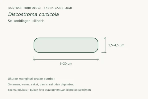
Saprobic on Quercus ilex L. Sexual morph: Illustrated in Wanasinghe et al. (2018). Asexual morph: Conidiomata (on host) 115–145 µm diam., 70–130 µm high, acervular, solitary to aggregated, semi-immersed, black; (on MEA) 50–70 µm diam., acervular, solitary to aggregated, erumpent, black. Conidiophores (on MEA) cylindrical, branched, hyaline, smooth, up to 30 µm long. Conidiogenous cells (on MEA) 7–18 × 2–3 µm (x– = 10.1 × 2.1 µm, n = 20) cylindrical, enteroblastic, annellidic, integrated or discrete, hyaline, determinate, smooth. Conidia (on MEA) 12–15 × (3–) 5–7 µm (x– = 13.5 × 5.4 µm, n = 47), obovoid, ellipsoid, broad fusiform or subcylindrical, straight or curved, hyaline when immature, pale to moderate brown at maturity, with 3 transverse, thick, darker septa, rarely constricted at the septa, often obtuse at both ends, or well rounded, smooth-walled, no appendage or sheath; basal cell obconic with a truncate base, pale brown or hyaline, thin-walled, 1–2.5 µm long (x– = 2 µm); two median cells doliiform, hyaline or pale brown, turning brown at maturity, together 5–7 µm long (x– = 6.1 µm), second cell from the base 1–3 µm long (x– = 2.5 µm), third cell from the base 1.4–4 µm long (x– = 2.6 µm); apical cell conical with obtuse or rounded apex, concolorous with the median cells, 1.8–3.5 µm long (x– = 2.5 µm). Colonies on MEA reaching 2–3 cm diam. after 11 days at 18 °C in darkness, filamentous, circular, flat with entire margin, white from above, reverse pale yellow.
Habitat dan peran ekologis
Saprobic on Quercus ilex L. Sexual morph: Illustrated in Wanasinghe et al. (2018). Asexual morph: Conidiomata (on host) 115–145 µm diam., 70–130 µm high, acervular, solitary to aggregated, semi-immersed, black; (on MEA) 50–70 µm diam., acervular, solitary to aggregated, erumpent, black. Conidiophores (on MEA) cylindrical, branched, hyaline, smooth, up to 30 µm long. Conidiogenous cells (on MEA) 7–18 × 2–3 µm (x– = 10.1 × 2.1 µm, n = 20) cylindrical, enteroblastic, annellidic, integrated or discrete, hyaline, determinate, smooth. Conidia (on MEA) 12–15 × (3–) 5–7 µm (x– = 13.5 × 5.4 µm, n = 47), obovoid, ellipsoid, broad fusiform or subcylindrical, straight or curved, hyaline when immature, pale to moderate brown at maturity, with 3 transverse, thick, darker septa, rarely constricted at the septa, often obtuse at both ends, or well rounded, smooth-walled, no appendage or sheath; basal cell obconic with a truncate base, pale brown or hyaline, thin-walled, 1–2.5 µm long (x– = 2 µm); two median cells doliiform, hyaline or pale brown, turning brown at maturity, together 5–7 µm long (x– = 6.1 µm), second cell from the base 1–3 µm long (x– = 2.5 µm), third cell from the base 1.4–4 µm long (x– = 2.6 µm); apical cell conical with obtuse or rounded apex, concolorous with the median cells, 1.8–3.5 µm long (x– = 2.5 µm).
- Habitat atau konteks temuan
- Informasi belum tersedia pada sumber rilis ini.
- Substrat
- Informasi belum tersedia pada sumber rilis ini.
- Peran atau konteks
- Pengurai
Reproduksi
Saprobic on Quercus ilex L. Sexual morph: Illustrated in Wanasinghe et al. (2018). Asexual morph: Conidiomata (on host) 115–145 µm diam., 70–130 µm high, acervular, solitary to aggregated, semi-immersed, black; (on MEA) 50–70 µm diam., acervular, solitary to aggregated, erumpent, black. Conidiophores (on MEA) cylindrical, branched, hyaline, smooth, up to 30 µm long. Conidiogenous cells (on MEA) 7–18 × 2–3 µm (x– = 10.1 × 2.1 µm, n = 20) cylindrical, enteroblastic, annellidic, integrated or discrete, hyaline, determinate, smooth. Conidia (on MEA) 12–15 × (3–) 5–7 µm (x– = 13.5 × 5.4 µm, n = 47), obovoid, ellipsoid, broad fusiform or subcylindrical, straight or curved, hyaline when immature, pale to moderate brown at maturity, with 3 transverse, thick, darker septa, rarely constricted at the septa, often obtuse at both ends, or well rounded, smooth-walled, no appendage or sheath; basal cell obconic with a truncate base, pale brown or hyaline, thin-walled, 1–2.5 µm long (x– = 2 µm); two median cells doliiform, hyaline or pale brown, turning brown at maturity, together 5–7 µm long (x– = 6.1 µm), second cell from the base 1–3 µm long (x– = 2.5 µm), third cell from the base 1.4–4 µm long (x– = 2.6 µm); apical cell conical with obtuse or rounded apex, concolorous with the median cells, 1.8–3.5 µm long (x– = 2.5 µm).
Catatan identitas dan cakupan
- Nama diterima mengikuti Catalogue of Life Base2026-09-11; uraian mempertahankan konsep dan nama sumber aslinya.
- Preserve original treatment localities, hosts and substrate scope. Materials examined describe documented specimens, not the full global range.
- Impor dan terjemahan otomatis belum diperiksa editorial per profil. Struktur reproduktif yang disebutkan tidak membuktikan seluruh siklus hidup diketahui.
- Pengukuran pada uraian mengikuti spesimen, kultur, dan metode sumber; angka tidak diperluas ke semua populasi.
Catatan distribusi dalam narasi mengikuti sumbernya. Dokumentasi spesimen atau foto mengikuti catatan sumber. Bukti wilayah Indonesia dan penilaian konservasi tidak disimpulkan dari gambar.
Melihat dengan konteks
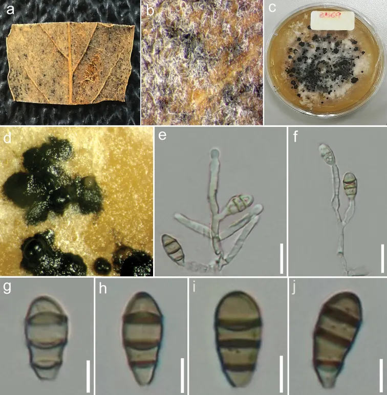
Sporocadus rosigena (MFLU 17-2803) a Leaf of Quercus ilex L b Close-up of conidiomata on host c Upper view of colony on MEA d Conidiomata in culture (MFLUCC 18-0387) e, f Different stages of conidiogenesis (MFLUCC 18-0387) g–j Conidia (MFLUCC 18-0387). Scale bars: 10 μm (e, f); 5 μm (g–j).
- Jenis
- Pelat ilmiah
- Kredit
- Digvijayini Bundhun, Rajesh Jeewon, Indunil C. Senanayake, Erio Camporesi, Janith V. S. Aluthmuhandiram, Alvin M. C. Tang, Ji-Chuan Kang, Vishwakalyan Bhoyroo, Kevin D. Hyde
- Lisensi
- CC BY 4.0
- Sumber media
- Buka entri sumber
Sumber profil
- [1] Morpho-molecular characterization of Discosia ravennica sp. nov. and a new host record for Sporocadus rosigena — Sporocadus rosigena
- [2] Catalogue of Life Base2026-09-11 — Sporocadus rosigena
- [3] Dokumentasi visual — Sporocadus rosigena
Rujukan halaman ini
Fungi. Sporocadus rosigena. Rilis data fungi-2026-10-12-expansion-2. https://fungi-atlas.mgadib.chatgpt.site/jamur/sporocadus-rosigena/.
Status: impor sumber; pemeriksaan editorial per profil belum selesai. Untuk klaim biologis, gunakan rujukan pada bagian terkait dan periksa konteks sumber aslinya.