01 / Morfologi

Ciri dan struktur

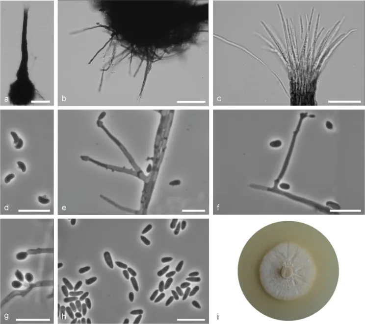
Sexual and asexual structures produced on sterilised beech twigs on surface of malt agar in Petri dishes. Ascomata abundant, superficially or partly embedded in the agar, single or in groups; ascomatal bases black, globose, (66–)89–153(–245) μm diam., with brown hyphal hairs, 12 to 165 μm long and 1 to 1.8 μm wide at the base; ascomatal necks black, straight or curved, (187–)272–462(–611) μm long, diameter (9–)10.4–16.7(22.5) μm at the apex and (26.8–)29.9–50.5(–63.9) μm at the base. Ostiolar hyphae present, pale brown, septate, straight or slightly waved, tapering towards the apex or sporadically dichotomous branching at the tip, (7–)8–16(–22) in number (17.8–)29.6–48.4(–64.5) μm long, (0.3–)0.5–1(–1.5) μm at the apex and (1.2–)1.6–2.3–(3) μm at the base. Asci evanescent. Ascospores one-celled, allantoid in side view (2.8–)3.1–3.8(–5.1) × (1–)1.1–1.4(–1.6) μm, elliptical in front view (2.8–)3.1–4.2(–4.8) × (1–)1.2–1.5(–1.8) μm, sometimes with residual sheath up to 1 μm thick, accumulated in creamy-colored mass at the tip of the neck. Conidiophores hyaline, micronematous, simple or branched, straight, simple or branched, bearing several conidiogenous cells, either borne on vegetative hyphae or on upright hyphae. Conidiogenous cells blastics, cylindrical, terminal, lateral or intercalary, straight or curved, tapering towards the apex, swollen apical part forming conidia by sympodial proliferation on visible denticles, (4.2–)17.5–43.1(–72.2) μm long, (0.8–)1.1–1.7(–2.1) μm wide at the base. Apical part with denticles (0.8–)1.3–3.7(–7.3) μm long and (1.2–)1.7–3.7(–7.3) μm wide.

Sebagian uraian ditampilkan dalam bahasa sumber; terjemahannya belum tersedia atau belum lolos pemeriksaan.

Rujukan: [1]

Pengukuran mengikuti objek, spesimen atau kultur, dan metode yang disebutkan sumber. Baca uraian asli untuk konteks lengkap.

02 / Kehidupan

Habitat dan peran ekologis

Sporothrix cracoviensis is phylogenetically distinct from the other Sporothrix species based on the βT, CAL and TEF1-α sequences. This species is closely related to S. fusiformis, S. lunata and S. prolifera. Sporothrix cracoviensis has smaller ascomatal necks (187–611 μm) compared to S. fusiformis (301–1168) μm (Aghayeva et al. 2004). Their conidial dimensions and shapes showed also differences. Sporothrix fusiforme has fusiforme conidia (Aghayeva et al. 2004), whereas S. cracoviensis has obovoid to clavate conidia. Sporothix lunata has also different shape of conidia (crescent) (Aghayeva et al. 2004) compared to S. cracoviensis (obovoid to clavate). In addition, S. lunata has smaller conidia (2.3–6.2 × 0.8–1.6 μm) (Aghayeva et al. 2004) compared to S. cracoviensis (2.8–8.7 μm × 1.1–2.7 μm). Sporothrix prolifera could be distinguished from S. cracoviensis by its smaller ascomatal base (S. prolifera: 65–90 μm (Kowalski and Butin 1989); S. cracoviensis: 66–245 μm) and smaller ascomatal necks (S. prolifera: 75–160 μm (Kowalski and Butin 1989); S. cracoviensis: 187–611 μm). In addition, S. prolifera has shorter ostiolar hyphae (S. prolifera: 15–30 μm (Kowalski and Butin 1989); S. cracoviensis: 26.8–63.9 μm) and shorter and wider ascospores (S. prolifera: 3.2–3.8 × 1.8–2 μm (Kowalski and Butin 1989); S. cracoviensis: 2.8–5.1 × 1–1.6 μm).

Sebagian uraian ditampilkan dalam bahasa sumber; terjemahannya belum tersedia atau belum lolos pemeriksaan.

Rujukan: [1]

Habitat atau konteks temuan
Informasi belum tersedia pada sumber rilis ini.
Substrat
Informasi belum tersedia pada sumber rilis ini.
Peran atau konteks
Belum dikategorikan pada rilis ini.
Temukan hubungan serupa
03 / Siklus hidup

Reproduksi

Sexual and asexual structures produced on sterilised beech twigs on surface of malt agar in Petri dishes. Ascomata abundant, superficially or partly embedded in the agar, single or in groups; ascomatal bases black, globose, (66–)89–153(–245) μm diam., with brown hyphal hairs, 12 to 165 μm long and 1 to 1.8 μm wide at the base; ascomatal necks black, straight or curved, (187–)272–462(–611) μm long, diameter (9–)10.4–16.7(22.5) μm at the apex and (26.8–)29.9–50.5(–63.9) μm at the base. Ostiolar hyphae present, pale brown, septate, straight or slightly waved, tapering towards the apex or sporadically dichotomous branching at the tip, (7–)8–16(–22) in number (17.8–)29.6–48.4(–64.5) μm long, (0.3–)0.5–1(–1.5) μm at the apex and (1.2–)1.6–2.3–(3) μm at the base.

Sebagian uraian ditampilkan dalam bahasa sumber; terjemahannya belum tersedia atau belum lolos pemeriksaan.

Rujukan: [1]

04 / Batas informasi

Catatan identitas dan cakupan

  • Nama diterima mengikuti Catalogue of Life Base2026-09-11; uraian mempertahankan konsep dan nama sumber aslinya.
  • Preserve original treatment localities, hosts and substrate scope. Materials examined describe documented specimens, not the full global range.
  • Impor dan terjemahan otomatis belum diperiksa editorial per profil. Struktur reproduktif yang disebutkan tidak membuktikan seluruh siklus hidup diketahui.
  • Pengukuran pada uraian mengikuti spesimen, kultur, dan metode sumber; angka tidak diperluas ke semua populasi.

Catatan distribusi dalam narasi mengikuti sumbernya. Dokumentasi spesimen atau foto mengikuti catatan sumber. Bukti wilayah Indonesia dan penilaian konservasi tidak disimpulkan dari gambar.

05 / Dokumentasi

Melihat dengan konteks

Sporothrix cracoviensis sp. nov. (CBS 147942) a ascoma b ascomatal base c ostiolar hyphae d ascospores e, f conidiogenous cell with an inflated cluster of denticles at the apex g conidiogenous cells arising directly from hyphae h conidia i fourteen-day-old culture on MEA. Scale bars: 50 μm (a, b), 25 μm (c), 10 μm (d–h).

Jenis
Pelat ilmiah
Kredit
Agnieszka Ostafińska, Robert Jankowiak, Piotr Bilański, Halvor Solheim, Michael J. Wingfield
Lisensi
CC BY 4.0
Sumber media
Buka entri sumber

Gambar menunjukkan objek menurut identifikasi sumber, bukan verifikasi independen spesimen oleh Fungi. Tidak ada skala baru yang ditambahkan.

06 / Rujukan

Sumber profil

  1. [1] Six new species of Sporothrix from hardwood trees in Poland — Sporothrix cracoviensisBagian sumber: PMC8357686_section_9
    Tanggal penghimpunan sumber: 2026-10-12
    Teks sumber: CC BY 4.0 · Ostafińska A, Jankowiak R, Bilański P, Solheim H, Wingfield MJ.
  2. [2] Catalogue of Life Base2026-09-11 — Sporothrix cracoviensisBagian sumber: ColDP NameUsage.tsv ID B2MD7; accepted species; source 2073
    Tanggal penghimpunan sumber: 2026-10-12
  3. [3] Dokumentasi visual — Sporothrix cracoviensisBagian sumber: Individual media record; identification follows the source, not independent determination
    Tanggal penghimpunan sumber: 2026-10-12
    Media sumber: CC BY 4.0 · Agnieszka Ostafińska, Robert Jankowiak, Piotr Bilański, Halvor Solheim, Michael J. Wingfield

Adaptasi teks: CC BY 4.0. Ostafińska A, Jankowiak R, Bilański P, Solheim H, Wingfield MJ. — Six new species of Sporothrix from hardwood trees in Poland — Sporothrix cracoviensis

Uraian asli dan riwayat sumber

Riwayat atau halaman sumber

Ringkasan sumber

Sexual and asexual structures produced on sterilised beech twigs on surface of malt agar in Petri dishes. Ascomata abundant, superficially or partly embedded in the agar, single or in groups; ascomatal bases black, globose, (66–)89–153(–245) μm diam., with brown hyphal hairs, 12 to 165 μm long and 1 to 1.8 μm wide at the base; ascomatal necks black, straight or curved, (187–)272–462(–611) μm long, diameter (9–)10.4–16.7(22.5) μm at the apex and (26.8–)29.9–50.5(–63.9) μm at the base. Ostiolar hyphae present, pale brown, septate, straight or slightly waved, tapering towards the apex or sporadically dichotomous branching at the tip, (7–)8–16(–22) in number (17.8–)29.6–48.4(–64.5) μm long, (0.3–)0.5–1(–1.5) μm at the apex and (1.2–)1.6–2.3–(3) μm at the base.

Revisi sumber

Morfologi sumber

Sexual and asexual structures produced on sterilised beech twigs on surface of malt agar in Petri dishes. Ascomata abundant, superficially or partly embedded in the agar, single or in groups; ascomatal bases black, globose, (66–)89–153(–245) μm diam., with brown hyphal hairs, 12 to 165 μm long and 1 to 1.8 μm wide at the base; ascomatal necks black, straight or curved, (187–)272–462(–611) μm long, diameter (9–)10.4–16.7(22.5) μm at the apex and (26.8–)29.9–50.5(–63.9) μm at the base. Ostiolar hyphae present, pale brown, septate, straight or slightly waved, tapering towards the apex or sporadically dichotomous branching at the tip, (7–)8–16(–22) in number (17.8–)29.6–48.4(–64.5) μm long, (0.3–)0.5–1(–1.5) μm at the apex and (1.2–)1.6–2.3–(3) μm at the base. Asci evanescent. Ascospores one-celled, allantoid in side view (2.8–)3.1–3.8(–5.1) × (1–)1.1–1.4(–1.6) μm, elliptical in front view (2.8–)3.1–4.2(–4.8) × (1–)1.2–1.5(–1.8) μm, sometimes with residual sheath up to 1 μm thick, accumulated in creamy-colored mass at the tip of the neck. Conidiophores hyaline, micronematous, simple or branched, straight, simple or branched, bearing several conidiogenous cells, either borne on vegetative hyphae or on upright hyphae. Conidiogenous cells blastics, cylindrical, terminal, lateral or intercalary, straight or curved, tapering towards the apex, swollen apical part forming conidia by sympodial proliferation on visible denticles, (4.2–)17.5–43.1(–72.2) μm long, (0.8–)1.1–1.7(–2.1) μm wide at the base. Apical part with denticles (0.8–)1.3–3.7(–7.3) μm long and (1.2–)1.7–3.7(–7.3) μm wide.

Revisi sumber

Ekologi sumber

Sporothrix cracoviensis is phylogenetically distinct from the other Sporothrix species based on the βT, CAL and TEF1-α sequences. This species is closely related to S. fusiformis, S. lunata and S. prolifera. Sporothrix cracoviensis has smaller ascomatal necks (187–611 μm) compared to S. fusiformis (301–1168) μm (Aghayeva et al. 2004). Their conidial dimensions and shapes showed also differences. Sporothrix fusiforme has fusiforme conidia (Aghayeva et al. 2004), whereas S. cracoviensis has obovoid to clavate conidia. Sporothix lunata has also different shape of conidia (crescent) (Aghayeva et al. 2004) compared to S. cracoviensis (obovoid to clavate). In addition, S. lunata has smaller conidia (2.3–6.2 × 0.8–1.6 μm) (Aghayeva et al. 2004) compared to S. cracoviensis (2.8–8.7 μm × 1.1–2.7 μm). Sporothrix prolifera could be distinguished from S. cracoviensis by its smaller ascomatal base (S. prolifera: 65–90 μm (Kowalski and Butin 1989); S. cracoviensis: 66–245 μm) and smaller ascomatal necks (S. prolifera: 75–160 μm (Kowalski and Butin 1989); S. cracoviensis: 187–611 μm). In addition, S. prolifera has shorter ostiolar hyphae (S. prolifera: 15–30 μm (Kowalski and Butin 1989); S. cracoviensis: 26.8–63.9 μm) and shorter and wider ascospores (S. prolifera: 3.2–3.8 × 1.8–2 μm (Kowalski and Butin 1989); S. cracoviensis: 2.8–5.1 × 1–1.6 μm).

Revisi sumber

Struktur reproduksi sumber

Sexual and asexual structures produced on sterilised beech twigs on surface of malt agar in Petri dishes. Ascomata abundant, superficially or partly embedded in the agar, single or in groups; ascomatal bases black, globose, (66–)89–153(–245) μm diam., with brown hyphal hairs, 12 to 165 μm long and 1 to 1.8 μm wide at the base; ascomatal necks black, straight or curved, (187–)272–462(–611) μm long, diameter (9–)10.4–16.7(22.5) μm at the apex and (26.8–)29.9–50.5(–63.9) μm at the base. Ostiolar hyphae present, pale brown, septate, straight or slightly waved, tapering towards the apex or sporadically dichotomous branching at the tip, (7–)8–16(–22) in number (17.8–)29.6–48.4(–64.5) μm long, (0.3–)0.5–1(–1.5) μm at the apex and (1.2–)1.6–2.3–(3) μm at the base.

Revisi sumber

Rujukan halaman ini

Fungi. Sporothrix cracoviensis. Rilis data fungi-2026-10-12-expansion-2. https://fungi-atlas.mgadib.chatgpt.site/jamur/sporothrix-cracoviensis/.

Status: impor sumber; pemeriksaan editorial per profil belum selesai. Untuk klaim biologis, gunakan rujukan pada bagian terkait dan periksa konteks sumber aslinya.