01 / Morfologi

Ciri dan struktur

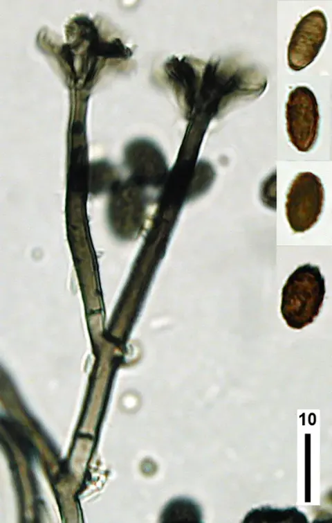
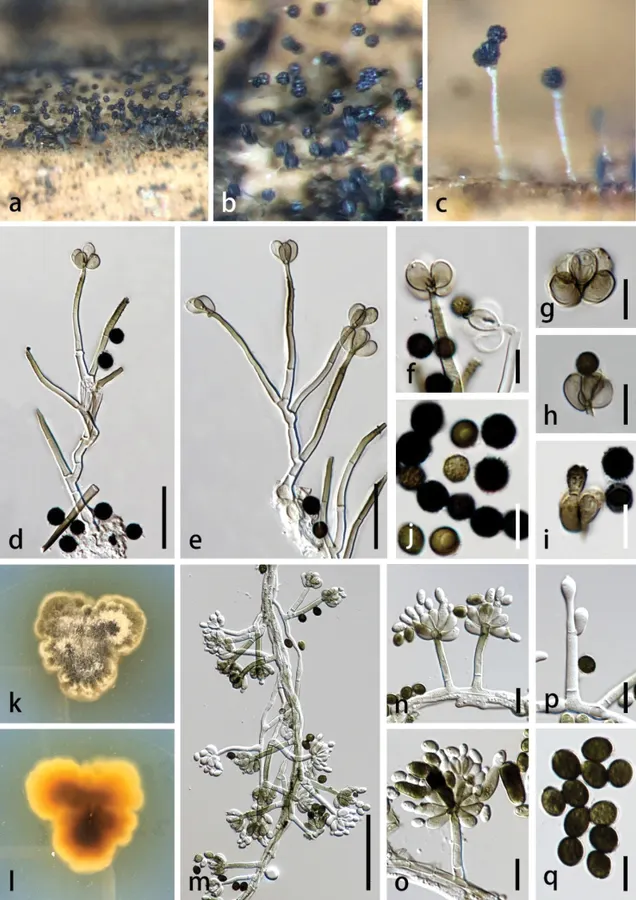
Saprobic on dead leaf of Agave sisalana. Sexual morph: undetermined. Asexual morph: Colonies superficial on host substrate, gregarious, visible as numerous black conidial masses. Conidiophores 35–70 × 2.5–5 µm (av. 48 × 4 μm, n = 30), macronematous, mononematous, irregularly or sympodially branched, straight or flexuous, subcylindrical, hyaline, becoming pale olivaceous brown in the above half, 1–3-septate, not constricted at the septa, smooth-walled, slightly rough-walled in the subterminal region, thick-walled, bearing 3–9 conidiogenous cells on the tip. Conidiogenous cells 6–10 × 4–6 µm (av. 8 × 5 μm, n = 30), enteroblastic, monophialidic, discrete, determinate, terminal, obovoid, sub-hyaline to pale olivaceous brown, smooth-walled. Conidia 5–7 µm diam. (av. 6 μm, n = 30), aggregating in slimy masses, globose, subglobose, aseptate, olivaceous brown to black, rough-walled, verrucose. Colonies on PDA reaching 2.5–3.0 cm in two weeks at 28 ± 2 °C, medium dense, raised, flat, floccose to fluffy, velvety, irregular edge, gold brown at the center, pale brown, with conidiophores forming on the surface of the medium, carrying slimy olivaceous green from above; brown to pale luteous from the reverse. The conidia producing on PDA after three weeks: Conidiophores 30–80 × 3.5–5.5 µm (av. 46 × 4 μm, n = 30), most similar with the above description, 0–2-septate, unbranched or branched, bearing 2–10 conidiogenous cells at the tip. Conidiogenous cells 9–17 × 4.5–7.5 µm (av. 12 × 5.5 μm, n = 30), most similar to those on natural substrate. Conidia 6.5–10 × 3–6.5 µm (av. 8 × 5 μm, n = 30), often aggregated as large, slimy, glistening, blackheads, initially hyaline to olivaceous green, oblong, obovoid to subglobose, becoming black-brown, globose, smooth to verruculose.

Sebagian uraian ditampilkan dalam bahasa sumber; terjemahannya belum tersedia atau belum lolos pemeriksaan.

Rujukan: [1]

Pengukuran mengikuti objek, spesimen atau kultur, dan metode yang disebutkan sumber. Baca uraian asli untuk konteks lengkap.

02 / Kehidupan

Habitat dan peran ekologis

• Canada, Cuba, India, Nigeria, and Pakistan (Ellis 1971; Jong and Davis 1976); • China (this study); Japan (Iwama et al. 2022); • New Guinea; Zaria (Jong and Davis 1976); • Thailand (Lin et al. 2016; Samarakoon et al. 2021); • Sudan (Lombard et al. 2016). Agave sisalana (this study), Arachishypogaea rhizosphere, soil (Jong and Davis 1976), Castanopsis cuspidata var. sieboldii (Iwama et al. 2022), dead plants, paper, seeds, and textiles (Ellis 1971), decaying shrubs, wood (Lin et al. 2016), Musa sp. (Samarakoon et al. 2021), soil in Mangifera field (Lombard et al. 2016).

Sebagian uraian ditampilkan dalam bahasa sumber; terjemahannya belum tersedia atau belum lolos pemeriksaan.

Rujukan: [1]

Habitat atau konteks temuan
Informasi belum tersedia pada sumber rilis ini.
Substrat
Informasi belum tersedia pada sumber rilis ini.
Peran atau konteks
Belum dikategorikan pada rilis ini.
Temukan hubungan serupa
03 / Siklus hidup

Reproduksi

Saprobic on dead leaf of Agave sisalana. Sexual morph: undetermined. Asexual morph: Colonies superficial on host substrate, gregarious, visible as numerous black conidial masses. Conidiophores 35–70 × 2.5–5 µm (av. 48 × 4 μm, n = 30), macronematous, mononematous, irregularly or sympodially branched, straight or flexuous, subcylindrical, hyaline, becoming pale olivaceous brown in the above half, 1–3-septate, not constricted at the septa, smooth-walled, slightly rough-walled in the subterminal region, thick-walled, bearing 3–9 conidiogenous cells on the tip. Conidiogenous cells 6–10 × 4–6 µm (av. 8 × 5 μm, n = 30), enteroblastic, monophialidic, discrete, determinate, terminal, obovoid, sub-hyaline to pale olivaceous brown, smooth-walled.

Sebagian uraian ditampilkan dalam bahasa sumber; terjemahannya belum tersedia atau belum lolos pemeriksaan.

Rujukan: [1]

Sinonim menurut acuan taksonomi rilis: Stachybotrys atrus var. microsporus
04 / Batas informasi

Catatan identitas dan cakupan

  • Nama diterima mengikuti Catalogue of Life Base2026-09-11; uraian mempertahankan konsep dan nama sumber aslinya.
  • Preserve original treatment localities, hosts and substrate scope. Materials examined describe documented specimens, not the full global range.
  • Impor dan terjemahan otomatis belum diperiksa editorial per profil. Struktur reproduktif yang disebutkan tidak membuktikan seluruh siklus hidup diketahui.
  • Pengukuran pada uraian mengikuti spesimen, kultur, dan metode sumber; angka tidak diperluas ke semua populasi.

Catatan distribusi dalam narasi mengikuti sumbernya. Dokumentasi spesimen atau foto mengikuti catatan sumber. Bukti wilayah Indonesia dan penilaian konservasi tidak disimpulkan dari gambar.

05 / Dokumentasi

Melihat dengan konteks

Stachybotrys microsporus (MHZU 23-0252, new host record) a–c colonies on the surface substrate d, e conidiophores f–i conidiogenous cells j conidia k colonies on pda (front) l colony on pda (below) m mycelium, conidiophores, and conidiogenous cells with conidia n–p conidiogenous cells with conidia q conidia. Scale bars: 20 µm (d, e); 50 µm (f–j, m); 10 µm (n–q).

Jenis
Pelat ilmiah
Kredit
Chunfang Liao, Mingkwan Doilom, D. Jayarama Bhat, Kandawatte Wedaralalage Thilini Chethana, Khanobporn Tangtrakulwanich, Yunhui Yang, Fatimah Al-Otibi, Kevin D. Hyde, Wei Dong
Lisensi
CC BY 4.0
Sumber media
Buka entri sumber

Gambar menunjukkan objek menurut identifikasi sumber, bukan verifikasi independen spesimen oleh Fungi. Tidak ada skala baru yang ditambahkan.

06 / Rujukan

Sumber profil

  1. [1] Unveiling four new taxa and Nigrosynnema natarajanensis comb. nov. in Stachybotryaceae (Hypocreales) from monocotyledon plants in Guangdong Province, China — Stachybotrys microsporusBagian sumber: PMC11905103_section_41
    Tanggal penghimpunan sumber: 2026-10-12
    Teks sumber: CC BY 4.0 · Liao C, Doilom M, Bhat DJ, Thilini Chethana KW, Tangtrakulwanich K, Yang Y, Al-Otibi F, Hyde KD, Dong W.
  2. [2] Catalogue of Life Base2026-09-11 — Stachybotrys microsporusBagian sumber: ColDP NameUsage.tsv ID 6ZF7K; accepted species; source 2073
    Tanggal penghimpunan sumber: 2026-10-12
  3. [3] Dokumentasi visual — Stachybotrys microsporusBagian sumber: Individual media record; identification follows the source, not independent determination
    Tanggal penghimpunan sumber: 2026-10-12
    Media sumber: CC BY 4.0 · Chunfang Liao, Mingkwan Doilom, D. Jayarama Bhat, Kandawatte Wedaralalage Thilini Chethana, Khanobporn Tangtrakulwanich, Yunhui Yang, Fatimah Al-Otibi, Kevin D. Hyde, Wei Dong

Adaptasi teks: CC BY 4.0. Liao C, Doilom M, Bhat DJ, Thilini Chethana KW, Tangtrakulwanich K, Yang Y, Al-Otibi F, Hyde KD, Dong W. — Unveiling four new taxa and Nigrosynnema natarajanensis comb. nov. in Stachybotryaceae (Hypocreales) from monocotyledon plants in Guangdong Province, China — Stachybotrys microsporus

Uraian asli dan riwayat sumber

Riwayat atau halaman sumber

Ringkasan sumber

Saprobic on dead leaf of Agave sisalana. Sexual morph: undetermined. Asexual morph: Colonies superficial on host substrate, gregarious, visible as numerous black conidial masses. Conidiophores 35–70 × 2.5–5 µm (av. 48 × 4 μm, n = 30), macronematous, mononematous, irregularly or sympodially branched, straight or flexuous, subcylindrical, hyaline, becoming pale olivaceous brown in the above half, 1–3-septate, not constricted at the septa, smooth-walled, slightly rough-walled in the subterminal region, thick-walled, bearing 3–9 conidiogenous cells on the tip.

Revisi sumber

Morfologi sumber

Saprobic on dead leaf of Agave sisalana. Sexual morph: undetermined. Asexual morph: Colonies superficial on host substrate, gregarious, visible as numerous black conidial masses. Conidiophores 35–70 × 2.5–5 µm (av. 48 × 4 μm, n = 30), macronematous, mononematous, irregularly or sympodially branched, straight or flexuous, subcylindrical, hyaline, becoming pale olivaceous brown in the above half, 1–3-septate, not constricted at the septa, smooth-walled, slightly rough-walled in the subterminal region, thick-walled, bearing 3–9 conidiogenous cells on the tip. Conidiogenous cells 6–10 × 4–6 µm (av. 8 × 5 μm, n = 30), enteroblastic, monophialidic, discrete, determinate, terminal, obovoid, sub-hyaline to pale olivaceous brown, smooth-walled. Conidia 5–7 µm diam. (av. 6 μm, n = 30), aggregating in slimy masses, globose, subglobose, aseptate, olivaceous brown to black, rough-walled, verrucose. Colonies on PDA reaching 2.5–3.0 cm in two weeks at 28 ± 2 °C, medium dense, raised, flat, floccose to fluffy, velvety, irregular edge, gold brown at the center, pale brown, with conidiophores forming on the surface of the medium, carrying slimy olivaceous green from above; brown to pale luteous from the reverse. The conidia producing on PDA after three weeks: Conidiophores 30–80 × 3.5–5.5 µm (av. 46 × 4 μm, n = 30), most similar with the above description, 0–2-septate, unbranched or branched, bearing 2–10 conidiogenous cells at the tip. Conidiogenous cells 9–17 × 4.5–7.5 µm (av. 12 × 5.5 μm, n = 30), most similar to those on natural substrate. Conidia 6.5–10 × 3–6.5 µm (av. 8 × 5 μm, n = 30), often aggregated as large, slimy, glistening, blackheads, initially hyaline to olivaceous green, oblong, obovoid to subglobose, becoming black-brown, globose, smooth to verruculose.

Revisi sumber

Ekologi sumber

• Canada, Cuba, India, Nigeria, and Pakistan (Ellis 1971; Jong and Davis 1976); • China (this study); Japan (Iwama et al. 2022); • New Guinea; Zaria (Jong and Davis 1976); • Thailand (Lin et al. 2016; Samarakoon et al. 2021); • Sudan (Lombard et al. 2016). Agave sisalana (this study), Arachishypogaea rhizosphere, soil (Jong and Davis 1976), Castanopsis cuspidata var. sieboldii (Iwama et al. 2022), dead plants, paper, seeds, and textiles (Ellis 1971), decaying shrubs, wood (Lin et al. 2016), Musa sp. (Samarakoon et al. 2021), soil in Mangifera field (Lombard et al. 2016).

Revisi sumber

Struktur reproduksi sumber

Saprobic on dead leaf of Agave sisalana. Sexual morph: undetermined. Asexual morph: Colonies superficial on host substrate, gregarious, visible as numerous black conidial masses. Conidiophores 35–70 × 2.5–5 µm (av. 48 × 4 μm, n = 30), macronematous, mononematous, irregularly or sympodially branched, straight or flexuous, subcylindrical, hyaline, becoming pale olivaceous brown in the above half, 1–3-septate, not constricted at the septa, smooth-walled, slightly rough-walled in the subterminal region, thick-walled, bearing 3–9 conidiogenous cells on the tip. Conidiogenous cells 6–10 × 4–6 µm (av. 8 × 5 μm, n = 30), enteroblastic, monophialidic, discrete, determinate, terminal, obovoid, sub-hyaline to pale olivaceous brown, smooth-walled.

Revisi sumber

Rujukan halaman ini

Fungi. Stachybotrys microsporus. Rilis data fungi-2026-10-12-expansion-2. https://fungi-atlas.mgadib.chatgpt.site/jamur/stachybotrys-microsporus/.

Status: impor sumber; pemeriksaan editorial per profil belum selesai. Untuk klaim biologis, gunakan rujukan pada bagian terkait dan periksa konteks sumber aslinya.