Ciri dan struktur
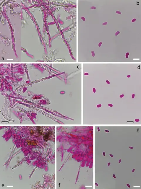
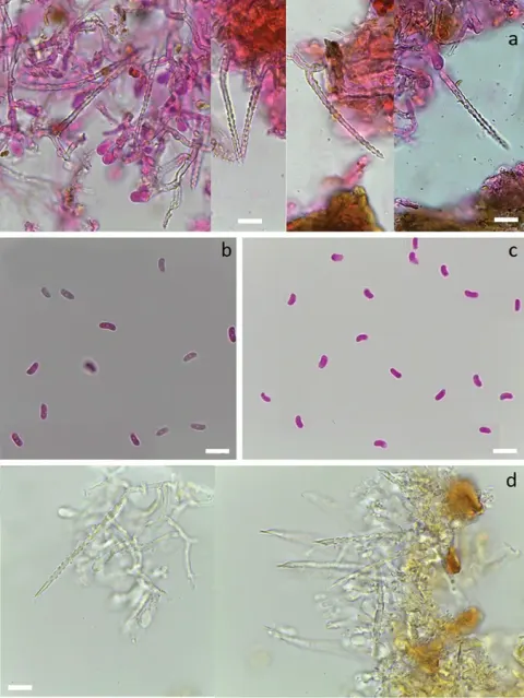
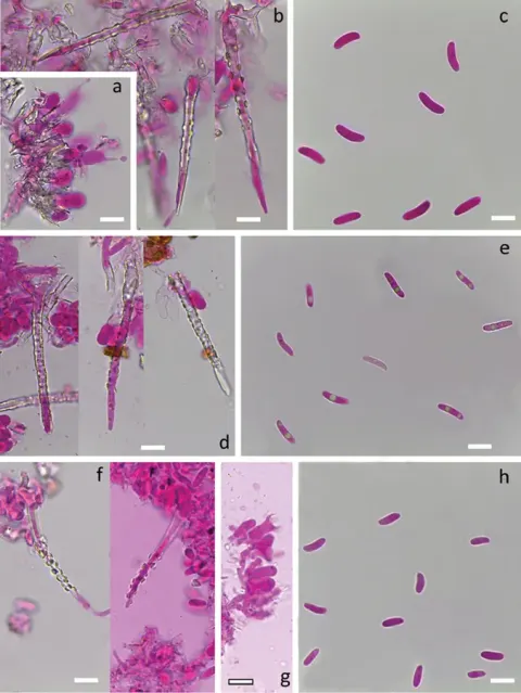
Hymenophore annual, membranous, adherent, effused forming small linear areas 5-15 x 1-2 cm; hymenial surface white, drying pallid cream, even, not creviced; margin thinning out, arachnoid, white, adherent. Context white, 100-200 µm thick, basal layer scanty, of a few parallel hyphae, intermediate layer of loosely arranged mainly ascending hyphae often branched at a wide angle, more freely in the subhymenium; generative hyphae 3-3-5 µm diameter, walls 0.2 µm thick, hyaline, with clamp connections, encrusted beneath the hymenium. Metuloids arising from the hymenium and hyphae of the intermediate layer, projecting to 60 µm, subulate with sometimes inflated bases, terminating in long-acuminate apices which often collapse, 60-112 x 4-5 µm, encrusted save at apices with flat tuberculate crystals, walls thinning towards apices. Hymenial layer to 30 µm deep, a lax palisade of basidia, paraphyses, and metuloids. Basidia subclavate, somewhat scanty, 20-26 x 5-6 µm, bearing 4 spores; sterigmata delicate, to 6 µm long. Paraphyses subclavate, somewhat scanty, 16-20 x 4-5 µm. Spores narrowly rod-shaped with rounded ends, sometimes vermiform or allantoid, 14-17 x 1.5-2.5 µm, walls smooth, hyaline, 0.2 µm thick.
Habitat dan peran ekologis
DISTRIBUTION: North Africa, Europe, Great Britain, North America, West Indies, New Zealand. HABITAT: Effused on bark or decorticated dead wood.
- Habitat atau konteks temuan
- Informasi belum tersedia pada sumber rilis ini.
- Substrat
- kulit kayu
- Peran atau konteks
- Belum dikategorikan pada rilis ini.
Reproduksi
Hymenophore annual, membranous, adherent, effused forming small linear areas 5-15 x 1-2 cm; hymenial surface white, drying pallid cream, even, not creviced; margin thinning out, arachnoid, white, adherent. Context white, 100-200 µm thick, basal layer scanty, of a few parallel hyphae, intermediate layer of loosely arranged mainly ascending hyphae often branched at a wide angle, more freely in the subhymenium; generative hyphae 3-3-5 µm diameter, walls 0.2 µm thick, hyaline, with clamp connections, encrusted beneath the hymenium. Metuloids arising from the hymenium and hyphae of the intermediate layer, projecting to 60 µm, subulate with sometimes inflated bases, terminating in long-acuminate apices which often collapse, 60-112 x 4-5 µm, encrusted save at apices with flat tuberculate crystals, walls thinning towards apices.
Catatan identitas dan cakupan
- Nama diterima mengikuti Catalogue of Life Base2026-09-11; uraian mempertahankan konsep dan nama sumber aslinya.
- Historical description as cited by BiotaNZ. Preserve explicit New Zealand/locality, host and specimen qualifiers. Distribution is the cited source scope, not a current global range.
- Impor dan terjemahan otomatis belum diperiksa editorial per profil. Struktur reproduktif yang disebutkan tidak membuktikan seluruh siklus hidup diketahui.
- Pengukuran pada uraian mengikuti spesimen, kultur, dan metode sumber; angka tidak diperluas ke semua populasi.
Catatan distribusi dalam narasi mengikuti sumbernya. Dokumentasi spesimen atau foto mengikuti catatan sumber. Bukti wilayah Indonesia dan penilaian konservasi tidak disimpulkan dari gambar.
Melihat dengan konteks
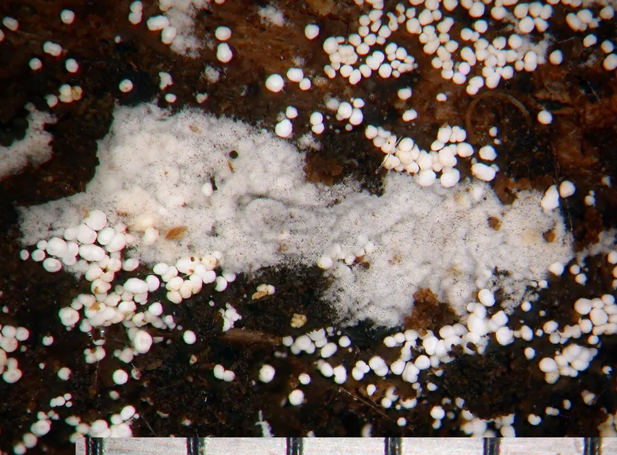
Dokumentasi Subulicystidium longisporum menurut asosiasi foto pada record takson iNaturalist. Kredit: Jerry Cooper. Identitas mengikuti sumber; bukan penentuan spesimen independen.
- Jenis
- Fotografi
- Kredit
- (c) Jerry Cooper, some rights reserved (CC BY), uploaded by Jerry Cooper
- Lisensi
- CC BY 4.0
- Sumber media
- Buka entri sumber
Dokumentasi tambahan
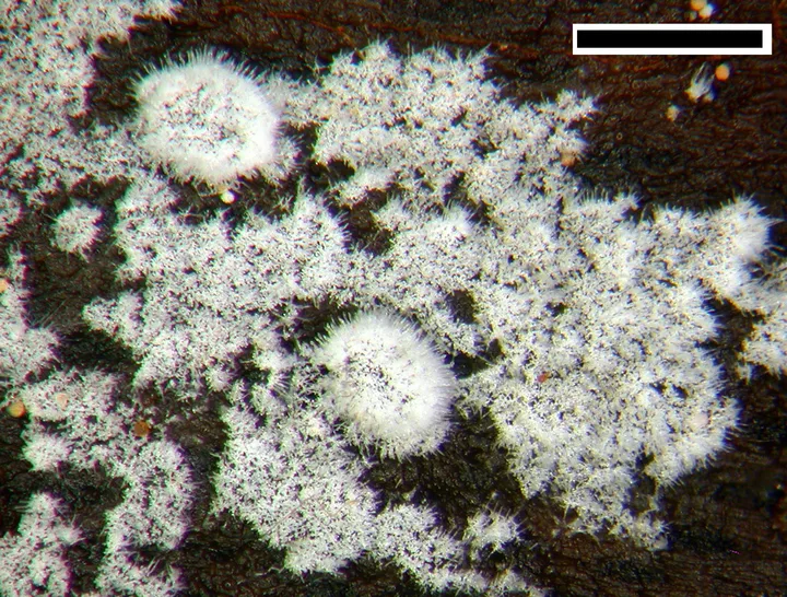
Foto yang diasosiasikan dengan Subulicystidium longisporum pada iNaturalist. Identifikasi mengikuti sumber; bukan penentuan spesimen independen.
Kredit dan penggunaan
(c) Jerry Cooper, some rights reserved (CC BY), uploaded by Jerry Cooper
Ukuran dan format gambar disesuaikan tanpa pemotongan, retus, atau penambahan skala. Lisensi sumber dipertahankan.
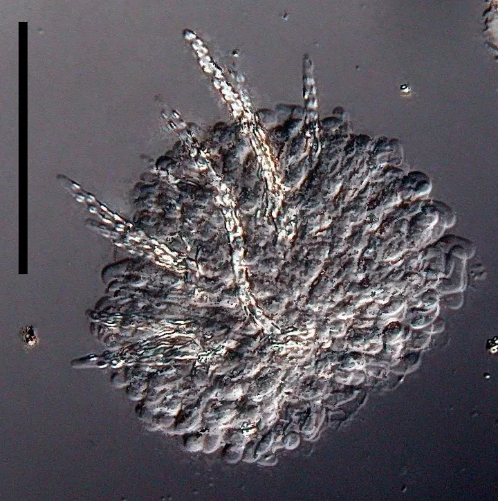
Foto yang diasosiasikan dengan Subulicystidium longisporum pada iNaturalist. Identifikasi mengikuti sumber; bukan penentuan spesimen independen.
Kredit dan penggunaan
(c) Jerry Cooper, some rights reserved (CC BY), uploaded by Jerry Cooper
Ukuran dan format gambar disesuaikan tanpa pemotongan, retus, atau penambahan skala. Lisensi sumber dipertahankan.
Sumber profil
- [1] Cunningham, G.H. 1963: The Thelephoraceae of Australia and New Zealand. New Zealand Department of Scientific and Industrial Research, Bulletin. 145. — Peniophora longispora
- [2] Catalogue of Life Base2026-09-11 — Subulicystidium longisporum
- [3] Dokumentasi visual — Subulicystidium longisporum
- [4] Dokumentasi visual — Subulicystidium longisporum
- [5] Dokumentasi visual — Subulicystidium longisporum
Rujukan halaman ini
Fungi. Subulicystidium longisporum. Rilis data fungi-2026-10-12-expansion-2. https://fungi-atlas.mgadib.chatgpt.site/jamur/subulicystidium-longisporum/.
Status: impor sumber; pemeriksaan editorial per profil belum selesai. Untuk klaim biologis, gunakan rujukan pada bagian terkait dan periksa konteks sumber aslinya.