Ciri dan struktur
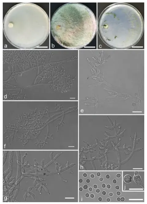
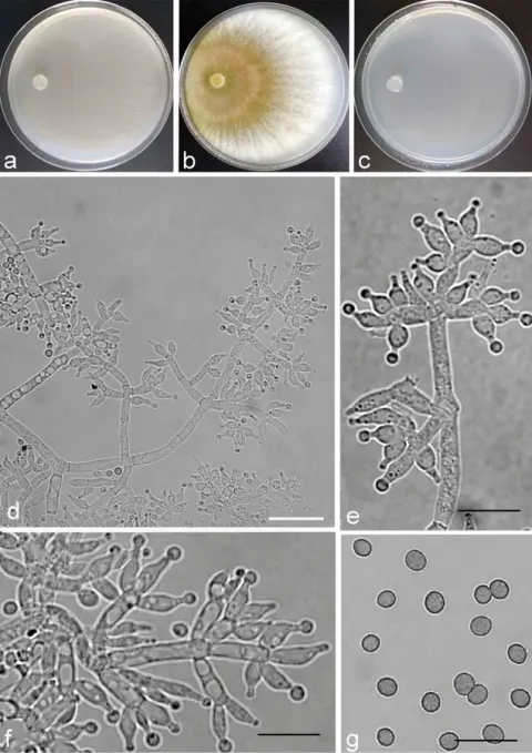
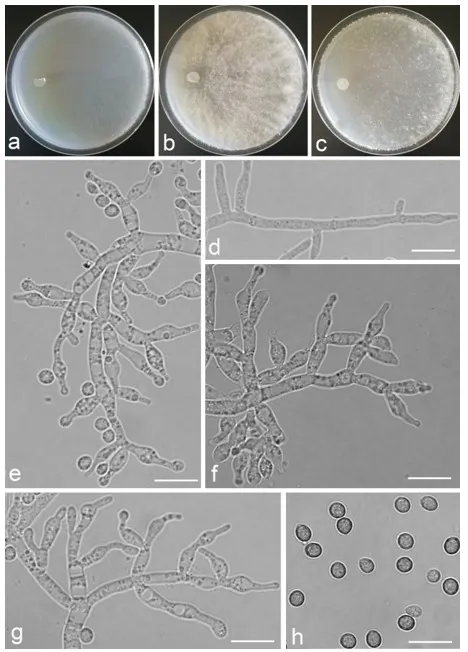
Culture characteristics: Optimum temperature for growth 25 °C. No growth at 35 °C in PDA and SNA. Colony radius on CMD after 72 h: 46–54 mm at 25 °C, 32–36 mm at 30 °C, 18–20 mm at 35 °C, covering the plate after 4 days at 25 °C. Colonies translucent, thin, flat, radial, mycelium loose. Aerial hyphae sparse, inconspicuous radiate. Chlamydospores unobserved. No diffusing pigment noted, odor indistinct. Colony radius on PDA after 72 h: 55–61 mm at 25 °C, 31–37 mm at 30 °C, covering the plate after 4 days at 25 °C. Colonies white, inconspicuous radial, mycelium dense. Aerial hyphae numerous, long and wooly. Chlamydospores unobserved. No diffusing pigment noted, odor indistinct. Colony radius on SNA after 72 h: 48–53 mm at 25 °C, 27–32 mm at 30 °C, covering the plate after 4 days at 25 °C. Colonies translucent, thin, radial. Aerial hyphae sparse, arachnoid, arranged in a scattered fashion. Pustules white, minute, abundant in the margin. Chlamydospores unobserved. No diffusing pigment noted, odor indistinct.
Habitat dan peran ekologis
Type: CHINA, Sichuan province, Daocheng county, N29°29′48.5″, E100°14′46.5″, 4362 m alt., endophytic on living stem of Hippuris vulgaris in pond, July 2014, Z.F. Yu, Y. Huang. Holotype YMF 1.04616, preserved in a metabolically inactive state (deep freezing) in the Conservation and Utilization of Bio-Resources in Yunnan. Ex-type culture CGMCC 3.19623. Culture characteristics: Optimum temperature for growth 25 °C. No growth at 35 °C in PDA and SNA. Colony radius on CMD after 72 h: 46–54 mm at 25 °C, 32–36 mm at 30 °C, 18–20 mm at 35 °C, covering the plate after 4 days at 25 °C. Colonies translucent, thin, flat, radial, mycelium loose. Aerial hyphae sparse, inconspicuous radiate. Chlamydospores unobserved. No diffusing pigment noted, odor indistinct. Colony radius on PDA after 72 h: 55–61 mm at 25 °C, 31–37 mm at 30 °C, covering the plate after 4 days at 25 °C. Colonies white, inconspicuous radial, mycelium dense. Aerial hyphae numerous, long and wooly. Chlamydospores unobserved. No diffusing pigment noted, odor indistinct. Colony radius on SNA after 72 h: 48–53 mm at 25 °C, 27–32 mm at 30 °C, covering the plate after 4 days at 25 °C. Colonies translucent, thin, radial. Aerial hyphae sparse, arachnoid, arranged in a scattered fashion.
- Habitat atau konteks temuan
- kultur yang didokumentasikan
- Substrat
- Informasi belum tersedia pada sumber rilis ini.
- Peran atau konteks
- Belum dikategorikan pada rilis ini.
Reproduksi
Notes: T richoderma scorpioideum is phylogenetically closely related to two species: T. viridescens and T. sempervirentis Jaklitsch & Voglmayr, the branches of T. scorpioideum and T. viridescens are slightly curved, whereas the branches of T. viridescens is paired and often terminated in 1 or 2 phialides [ 59 ], which distinctly differs from verticillate branches of T. scorpioideum. As for the phialides of T. viridescens, it often forms a submoniliform chain of 2–6 cells when cultured on CMD [ 59 ], in contrast, the structure is inconspicuous on the phialides of T. scorpioideum. Furthermore, the conidia of T. viridescens sometimes are wider (3.0–3.7 μm) than those of T. scorpioideum.
Catatan identitas dan cakupan
- Nama diterima mengikuti Catalogue of Life Base2026-09-11; uraian mempertahankan konsep dan nama sumber aslinya.
- Preserve original treatment localities, hosts and substrate scope. Materials examined describe documented specimens, not the full global range.
- Impor dan terjemahan otomatis belum diperiksa editorial per profil. Struktur reproduktif yang disebutkan tidak membuktikan seluruh siklus hidup diketahui.
- Pengukuran pada uraian mengikuti spesimen, kultur, dan metode sumber; angka tidak diperluas ke semua populasi.
Catatan distribusi dalam narasi mengikuti sumbernya. Dokumentasi spesimen atau foto mengikuti catatan sumber. Bukti wilayah Indonesia dan penilaian konservasi tidak disimpulkan dari gambar.
Melihat dengan konteks
Trichoderma scorpioideum (YMF 1.04616). (a – c) Cultures after 7 d at 25 °C (a) on CMD; (b) on PDA; (c) on SNA). (d) Conidiophores formed on SNA. (e – g) Conidiophores, phialides and conidia formed on SNA. (h) Conidia. Scale bars: (d – h) = 10 μm.
- Jenis
- Pelat ilmiah
- Kredit
- © 2021 by the authors.
- Lisensi
- CC BY 4.0
- Sumber media
- Buka entri sumber
Sumber profil
- [1] New Species of Trichoderma Isolated as Endophytes and Saprobes from Southwest China — Trichoderma scorpioideum
- [2] Catalogue of Life Base2026-09-11 — Trichoderma scorpioideum
- [3] Dokumentasi visual — Trichoderma scorpioideum
Rujukan halaman ini
Fungi. Trichoderma scorpioideum. Rilis data fungi-2026-10-12-expansion-2. https://fungi-atlas.mgadib.chatgpt.site/jamur/trichoderma-scorpioideum/.
Status: impor sumber; pemeriksaan editorial per profil belum selesai. Untuk klaim biologis, gunakan rujukan pada bagian terkait dan periksa konteks sumber aslinya.