Ciri dan struktur
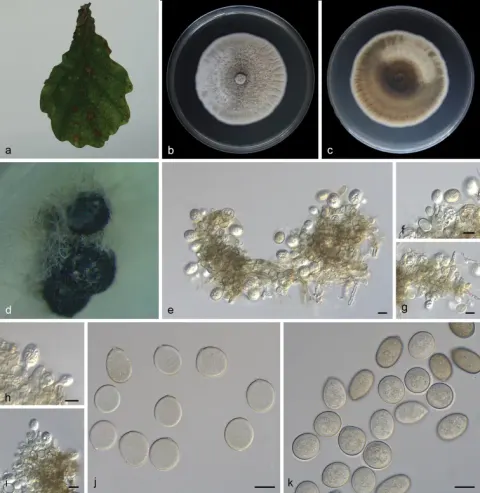
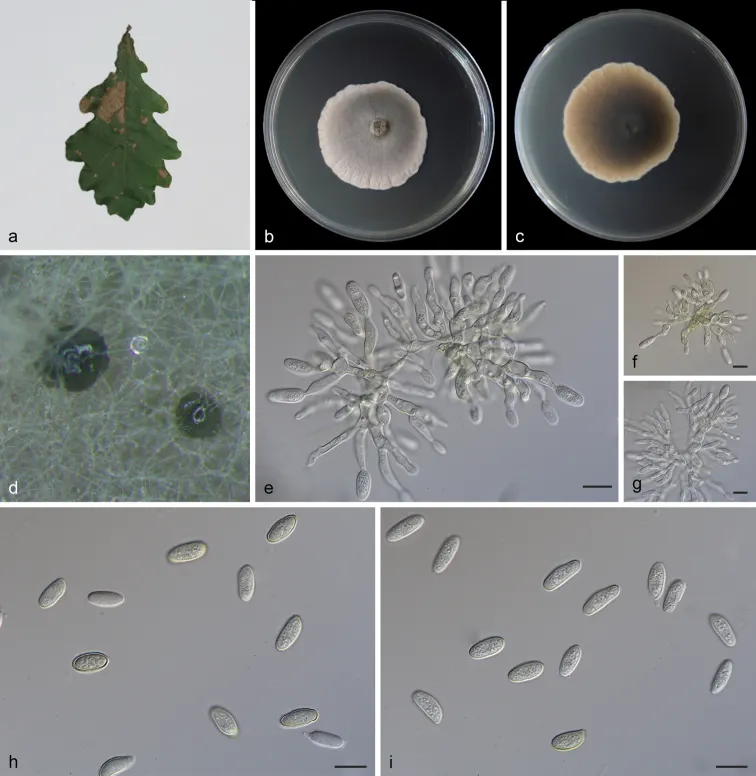
Asexual morph: Living as endophyte in leaves, forming distinct leaf lesions, shape and size variable, subcircular to angular-irregular, pale brown to brown. Colonies on PDA incubated at 25°C in the dark with an average radial growth rate of 5–7 mm/d and occupying an entire 90 mm Petri dish in 14 d, forming some conspicuous concentric circles, aerial mycelium cottony, white initially, then becoming greyish-sepia. Conidiomata sporodochial, appeared within 14 days or longer, formed on agar surface, slimy, black, semi-submerged. Sporodochial conidiophores densely and irregularly branched, 11.0–24.0 μm × 1.5–5.0 μm, bearing apical whorls of 2–3 phialides; sporodochial phialides monophialidic, subulate to subcylindrical, 9.0–16.0 μm × 1.5–5.0 μm, smooth, thin-walled, apex obtuse to truncate, sometimes forming indistinct periclinal thickenings. Conidia solitary, ellipsoid to obovoid, 6.5–14.0 μm × 4.0–6.0 μm, wall thin, up to 1.0 μm, hyaline to subhyaline, smooth, apex and base broadly rounded, with inconspicuous to conspicuous basal hilum (frill), occasionally somewhat peg-like and truncate when conspicuous. Microconidia not observed. Sexual morph not observed. Cultures incubated on MEA at 25°C in darkness, attaining 38.0–42.0 mm diam. after 14 d (growth rate 2.7–3.0 mm diam./d), margin scalloped, at first creamy white, grey near the centre, reverse light brown to dark, with olivaceous edge. Conidial formation not observed.
Habitat dan peran ekologis
Asexual morph: Living as endophyte in leaves, forming distinct leaf lesions, shape and size variable, subcircular to angular-irregular, pale brown to brown. Colonies on PDA incubated at 25°C in the dark with an average radial growth rate of 5–7 mm/d and occupying an entire 90 mm Petri dish in 14 d, forming some conspicuous concentric circles, aerial mycelium cottony, white initially, then becoming greyish-sepia. Conidiomata sporodochial, appeared within 14 days or longer, formed on agar surface, slimy, black, semi-submerged. Sporodochial conidiophores densely and irregularly branched, 11.0–24.0 μm × 1.5–5.0 μm, bearing apical whorls of 2–3 phialides; sporodochial phialides monophialidic, subulate to subcylindrical, 9.0–16.0 μm × 1.5–5.0 μm, smooth, thin-walled, apex obtuse to truncate, sometimes forming indistinct periclinal thickenings. Conidia solitary, ellipsoid to obovoid, 6.5–14.0 μm × 4.0–6.0 μm, wall thin, up to 1.0 μm, hyaline to subhyaline, smooth, apex and base broadly rounded, with inconspicuous to conspicuous basal hilum (frill), occasionally somewhat peg-like and truncate when conspicuous. Microconidia not observed. Sexual morph not observed. Braun et al. (2018) described Tubakia dryinoides, based on morphological and molecular data.
- Habitat atau konteks temuan
- kultur yang didokumentasikan
- Substrat
- Informasi belum tersedia pada sumber rilis ini.
- Peran atau konteks
- Belum dikategorikan pada rilis ini.
Reproduksi
Asexual morph: Living as endophyte in leaves, forming distinct leaf lesions, shape and size variable, subcircular to angular-irregular, pale brown to brown. Colonies on PDA incubated at 25°C in the dark with an average radial growth rate of 5–7 mm/d and occupying an entire 90 mm Petri dish in 14 d, forming some conspicuous concentric circles, aerial mycelium cottony, white initially, then becoming greyish-sepia. Conidiomata sporodochial, appeared within 14 days or longer, formed on agar surface, slimy, black, semi-submerged. Sporodochial conidiophores densely and irregularly branched, 11.0–24.0 μm × 1.5–5.0 μm, bearing apical whorls of 2–3 phialides; sporodochial phialides monophialidic, subulate to subcylindrical, 9.0–16.0 μm × 1.5–5.0 μm, smooth, thin-walled, apex obtuse to truncate, sometimes forming indistinct periclinal thickenings.
Catatan identitas dan cakupan
- Nama diterima mengikuti Catalogue of Life Base2026-09-11; uraian mempertahankan konsep dan nama sumber aslinya.
- Preserve original treatment localities, hosts and substrate scope. Materials examined describe documented specimens, not the full global range.
- Impor dan terjemahan otomatis belum diperiksa editorial per profil. Struktur reproduktif yang disebutkan tidak membuktikan seluruh siklus hidup diketahui.
- Pengukuran pada uraian mengikuti spesimen, kultur, dan metode sumber; angka tidak diperluas ke semua populasi.
Catatan distribusi dalam narasi mengikuti sumbernya. Dokumentasi spesimen atau foto mengikuti catatan sumber. Bukti wilayah Indonesia dan penilaian konservasi tidak disimpulkan dari gambar.
Melihat dengan konteks
Tubakia dryinoides (SAUCC 1924). a diseased leaf of Quercus palustris; b surface of colony after 15 days on MEA; c reverse of colony after 15 days on MEA; d conidiomata; e–g conidiophores, conidiogenous cells with conidia; h–i conidia. Scale bars: 10 μm (e–i).
- Jenis
- Pelat ilmiah
- Kredit
- Zhaoxue Zhang, Taichang Mu, Shubin Liu, Rongyu Liu, Xiuguo Zhang, Jiwen Xia
- Lisensi
- CC BY 4.0
- Sumber media
- Buka entri sumber
Sumber profil
- [1] Morphological and phylogenetic analyses reveal a new genus and two new species of Tubakiaceae from China — Tubakia dryinoides
- [2] Catalogue of Life Base2026-09-11 — Tubakia dryinoides
- [3] Dokumentasi visual — Tubakia dryinoides
Rujukan halaman ini
Fungi. Tubakia dryinoides. Rilis data fungi-2026-10-12-expansion-2. https://fungi-atlas.mgadib.chatgpt.site/jamur/tubakia-dryinoides/.
Status: impor sumber; pemeriksaan editorial per profil belum selesai. Untuk klaim biologis, gunakan rujukan pada bagian terkait dan periksa konteks sumber aslinya.