Ciri dan struktur
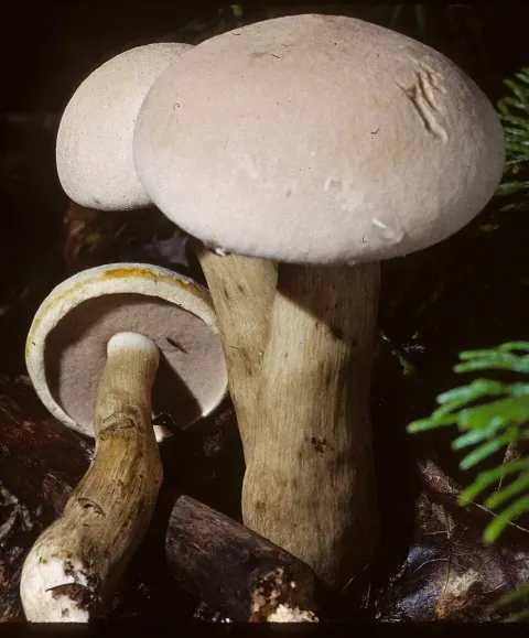
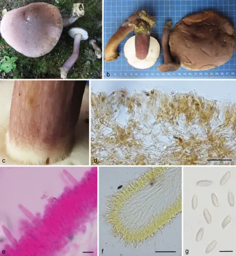
The cap of this species grows up to 15 cm (6 in) in diameter, and rarely infested by insects. The colour of the spore print is brownish with a reddish or pink tint. Spores are somewhat fuse-shaped, smooth, and measure 11–17 by 3–5 μm. The basidia (spore-bearing cells) are club-shaped, four-spored, and measure 18–25.6 by 7.0–10.2 μm. Cystidia on the walls of the tubes (pleurocystidia) are fuse-shaped with a central swelling, thin-walled, and have granular contents. They possess sharp to tapered tips, and have overall dimensions of 36–44 by 8.0–11.0 μm. On the pore edges, the cheilocystidia are similar in shape to the pleurocystidia, measuring 24.8–44.0 by 7.3–11.0 μm. The hymenium of Smith and Thiers's variety uliginosus, when mounted in Melzer's reagent, shows reddish globules of pigment measuring 2–8 μm that appear in the hyphae and throughout the hymenium, and a large (8–12 μm) globule in the pleurocystidia. Several chemical tests have been documented that can help confirm the identify of this species. On the cap flesh, application of formaldehyde turns the tissue pinkish, iron salts result in a colour change to greyish-green, aniline causes a lavender to reddish-brown colour, and phenol a purplish pink to reddish brown. On the cap cuticle, nitric acid causes an orange-salmon colour, sulphuric acid creates orange-red, ammonia usually makes brown, and a potassium hydroxide solution usually makes orange.
Habitat dan peran ekologis
Like all Tylopilus species, T. felleus is mycorrhizal. It is found in deciduous and coniferous woodland, often under beech and oak in well-drained acid soils, which can be sandy, gravelly or peaty. If encountered on calcareous (chalky) soil, it will be in moist areas that have become waterlogged and have ample leaf litter. Fruit bodies grow singly or in small groups, and occasionally in small clusters with two or three joined at the base of the stem. Fruit bodies have also been growing in the cavities of old trees, on old conifer stumps, or on buried rotten wood. The fungus obtains most of its nitrogen requirements from amino acids derived from the breakdown of proteins, although a lesser amount is obtained from the amino sugar glucosamine (a breakdown product of chitin, a major component of fungal cell walls). The mycorrhizal plant partner benefits from the fungus's ability to use these forms of nitrogen, which are often abundant in the forest floor. Fruit bodies appear over summer and autumn, anytime from June to October or even November, in many of the northern temperate zones. Large numbers may appear in some years and none in others, generally proportional to the amount of rainfall.
- Habitat atau konteks temuan
- hutan
- Substrat
- Informasi belum tersedia pada sumber rilis ini.
- Peran atau konteks
- Mitra tumbuhan
Reproduksi
The colour of the spore print is brownish with a reddish or pink tint. Spores are somewhat fuse-shaped, smooth, and measure 11–17 by 3–5 μm. The basidia (spore-bearing cells) are club-shaped, four-spored, and measure 18–25.6 by 7.0–10.2 μm. Cystidia on the walls of the tubes (pleurocystidia) are fuse-shaped with a central swelling, thin-walled, and have granular contents. They possess sharp to tapered tips, and have overall dimensions of 36–44 by 8.0–11.0 μm. On the pore edges, the cheilocystidia are similar in shape to the pleurocystidia, measuring 24.8–44.0 by 7.3–11.0 μm. The hymenium of Smith and Thiers's variety uliginosus, when mounted in Melzer's reagent, shows reddish globules of pigment measuring 2–8 μm that appear in the hyphae and throughout the hymenium, and a large (8–12 μm) globule in the pleurocystidia.
Catatan identitas dan cakupan
- Nama diterima mengikuti Catalogue of Life Base2026-09-11; uraian mempertahankan konsep dan nama sumber aslinya.
- Original geographic, specimen and life-cycle scope retained; source import awaits individual editorial review.
- Impor dan terjemahan otomatis belum diperiksa editorial per profil. Struktur reproduktif yang disebutkan tidak membuktikan seluruh siklus hidup diketahui.
- Pengukuran pada uraian mengikuti spesimen, kultur, dan metode sumber; angka tidak diperluas ke semua populasi.
Catatan distribusi dalam narasi mengikuti sumbernya. Dokumentasi spesimen atau foto mengikuti catatan sumber. Bukti wilayah Indonesia dan penilaian konservasi tidak disimpulkan dari gambar.
Melihat dengan konteks
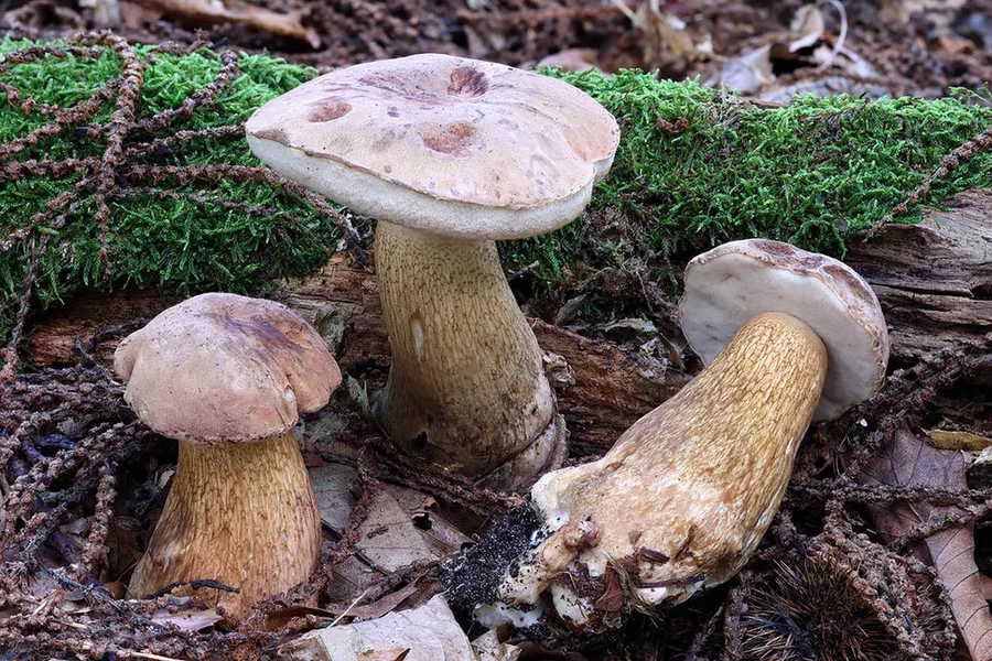
Dokumentasi Tylopilus felleus menurut asosiasi foto pada record takson iNaturalist. Kredit: Daniel Ghyselinck. Identitas mengikuti sumber; bukan penentuan spesimen independen.
- Jenis
- Fotografi
- Kredit
- (c) Daniel Ghyselinck, some rights reserved (CC BY), uploaded by Daniel Ghyselinck
- Lisensi
- CC BY 4.0
- Sumber media
- Buka entri sumber
Dokumentasi tambahan
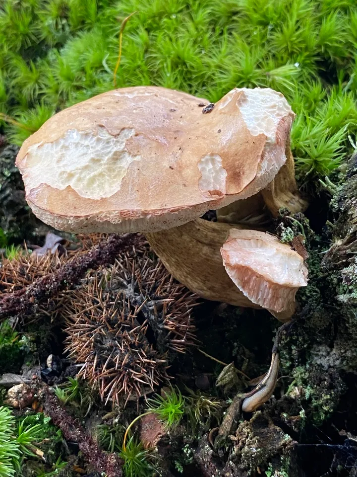
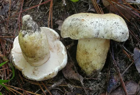
Foto yang diasosiasikan dengan Tylopilus felleus pada iNaturalist. Identifikasi mengikuti sumber; bukan penentuan spesimen independen.
Kredit dan penggunaan
(c) Thomas Dreux, some rights reserved (CC BY), uploaded by Thomas Dreux
Ukuran dan format gambar disesuaikan tanpa pemotongan, retus, atau penambahan skala. Lisensi sumber dipertahankan.
Sumber profil
- [1] Tylopilus felleus — Wikipedia (en)
- [2] Catalogue of Life Base2026-09-11 — Tylopilus felleus
- [3] Dokumentasi visual — Tylopilus felleus
- [4] Dokumentasi visual — Tylopilus felleus
Rujukan halaman ini
Fungi. Tylopilus felleus. Rilis data fungi-2026-10-12-expansion-2. https://fungi-atlas.mgadib.chatgpt.site/jamur/tylopilus-felleus/.
Status: impor sumber; pemeriksaan editorial per profil belum selesai. Untuk klaim biologis, gunakan rujukan pada bagian terkait dan periksa konteks sumber aslinya.