Ciri dan struktur
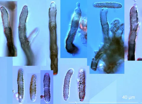
Zasmidium cellare growth typically includes hyphae (up to 5000 μm in length with walls 0.5-0.6 μm thick) upon which spores are produced at the end of teeth-like structures that are less pigmented than the basal portion of the conidiophore. Aerial hyphae are usually rough and darkly coloured, 2-2.5 μm wide, with thick walls, while the submerged hyphae are smooth and 2-3 μm wide. The Aerial hyphae are observed to have conidiogenous cells (thought to be remnants of conidiophores) primarily on the terminal of the hyphae, sometimes on the sides, that are cylindrical and 20-60 μm long and 2-2.5 μm wide. The structure of the colony is circular (7mm in diameter after 14 days at 24 °C in vitro), elevated and fuzzy in texture due to branched, filamentous growth. Large colonies can amalgamate into amorphous structures that optimizes the absorption of volatile compounds from the air, moving away from more circular shapes and creating sheets of mycelium, especially under very humid conditions. In vitro, Z. cellare is characterized as being morphologically similar to Stenella araguata, which both reside in the order Capnodiales and family Mycosphaerellaceae.
Habitat dan peran ekologis
Zasmidium cellare was first recorded existing is cellars in 1696. The presence of Z. cellare in any of these regions is completely dependent on human activity, however. For example, there are wineries in Italy that are devoid of Z. cellare but also maintain a level of cleanliness in their cellars, while other Italian wineries cherish its presence and often feed it left over wine, encouraging its proliferation. When Z. cellare is present, however, it is commonly found on brickwork and timber next to its food sources and can also, on the rare occasion, be found in nearby soil.
- Habitat atau konteks temuan
- Informasi belum tersedia pada sumber rilis ini.
- Substrat
- Informasi belum tersedia pada sumber rilis ini.
- Peran atau konteks
- Belum dikategorikan pada rilis ini.
Reproduksi
Zasmidium cellare growth typically includes hyphae (up to 5000 μm in length with walls 0.5-0.6 μm thick) upon which spores are produced at the end of teeth-like structures that are less pigmented than the basal portion of the conidiophore. Aerial hyphae are usually rough and darkly coloured, 2-2.5 μm wide, with thick walls, while the submerged hyphae are smooth and 2-3 μm wide. The Aerial hyphae are observed to have conidiogenous cells (thought to be remnants of conidiophores) primarily on the terminal of the hyphae, sometimes on the sides, that are cylindrical and 20-60 μm long and 2-2.5 μm wide. The structure of the colony is circular (7mm in diameter after 14 days at 24 °C in vitro), elevated and fuzzy in texture due to branched, filamentous growth.
Catatan identitas dan cakupan
- Nama diterima mengikuti Catalogue of Life Base2026-09-11; uraian mempertahankan konsep dan nama sumber aslinya.
- Original geographic, specimen and life-cycle scope retained; source import awaits individual editorial review.
- Impor dan terjemahan otomatis belum diperiksa editorial per profil. Struktur reproduktif yang disebutkan tidak membuktikan seluruh siklus hidup diketahui.
- Pengukuran pada uraian mengikuti spesimen, kultur, dan metode sumber; angka tidak diperluas ke semua populasi.
Catatan distribusi dalam narasi mengikuti sumbernya. Dokumentasi spesimen atau foto mengikuti catatan sumber. Bukti wilayah Indonesia dan penilaian konservasi tidak disimpulkan dari gambar.
Melihat dengan konteks
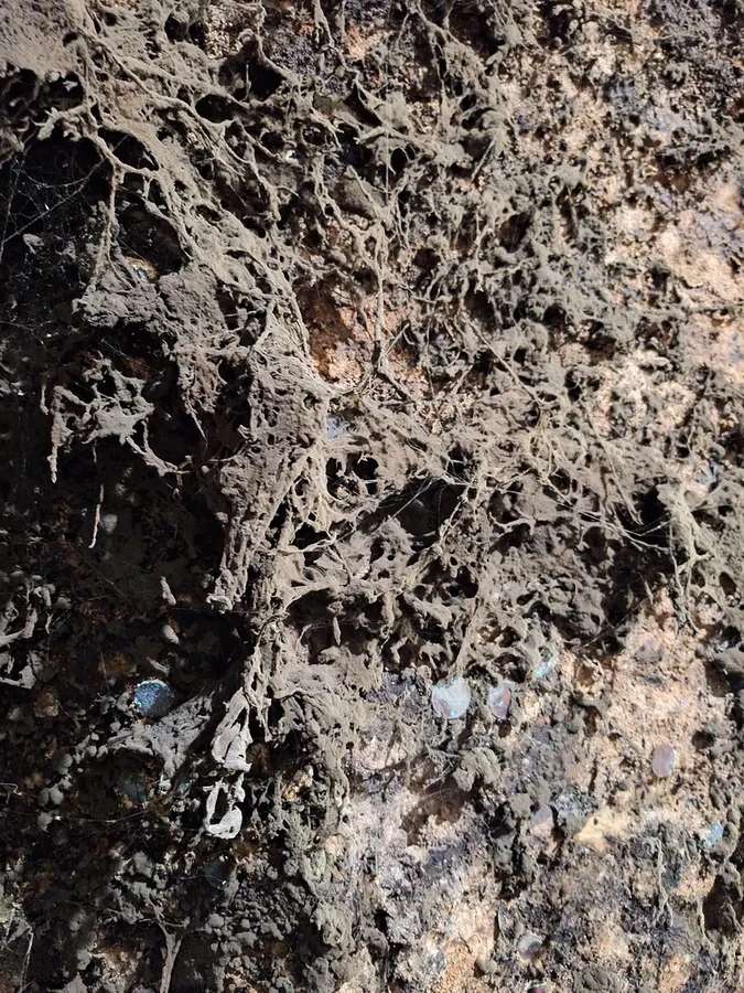
Dokumentasi Zasmidium cellare menurut asosiasi foto pada record takson iNaturalist. Kredit: Dávid Horváth. Identitas mengikuti sumber; bukan penentuan spesimen independen.
- Jenis
- Fotografi
- Kredit
- (c) Dávid Horváth, some rights reserved (CC BY), uploaded by Dávid Horváth
- Lisensi
- CC BY 4.0
- Sumber media
- Buka entri sumber
Sumber profil
- [1] Zasmidium cellare — Wikipedia (en)
- [2] Catalogue of Life Base2026-09-11 — Zasmidium cellare
- [3] Dokumentasi visual — Zasmidium cellare
Rujukan halaman ini
Fungi. Zasmidium cellare. Rilis data fungi-2026-10-12-expansion-2. https://fungi-atlas.mgadib.chatgpt.site/jamur/zasmidium-cellare/.
Status: impor sumber; pemeriksaan editorial per profil belum selesai. Untuk klaim biologis, gunakan rujukan pada bagian terkait dan periksa konteks sumber aslinya.