Ciri dan struktur
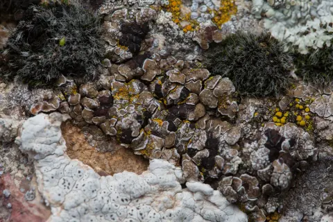
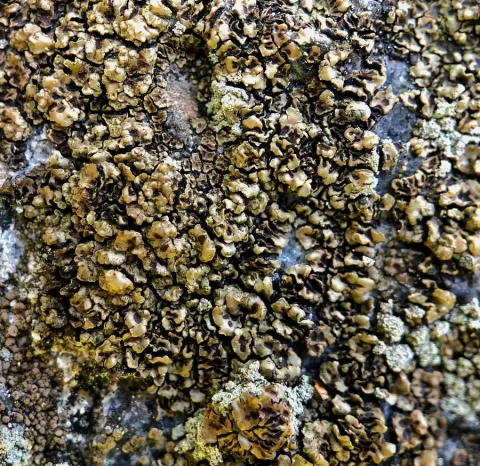
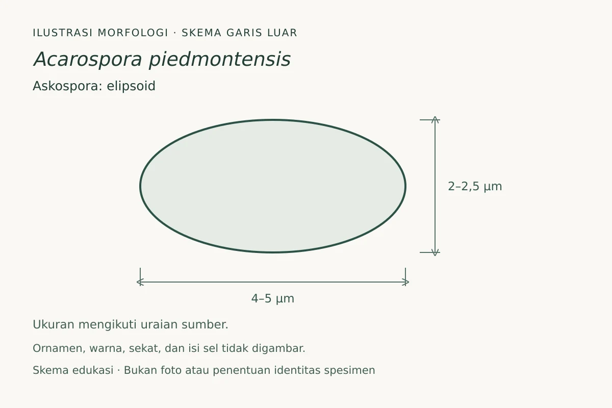
The thallus (lichen body) begins as scattered areoles (small surface units) that develop into small squamules (scale-like lobes), sometimes merging into a more continuous patch as the thallus expands. Squamules are typically about 0.5–1 mm across and less than 1 mm tall, and each develops a stalk (stipe) that is narrower than the squamule itself. The upper surface is brown and matte, and lacks pruina (a powdery coating), with the edges turned downward, often with a black rim. The lower surface is brown but commonly becomes blackened (melanized, darkened by pigment), and it may show deep fissures. There is usually one apothecium per squamule. The disc is immersed (sunken into the squamule) and dull brown, becoming red-brown when wet, and can reach about 1 mm in diameter. Under the microscope, the is about 20–30 μm (micrometres) thick, with a dark upper layer and a hyaline (colorless) lower portion, and the can be thin to uneven, with the (the photosynthetic partner) often arranged in vertical rows. The hymenium (spore-bearing layer) in mature apothecia is reported as (100–)150–170 μm tall, and the ascospores are single-celled (), hyaline, broadly ellipsoid, about 4–5 × 2–2.5 μm. Pycnidia were observed (about 80 μm diameter), with conidia about 3 × 1 μm. No secondary metabolites were detected by thin-layer chromatography; standard lichen spot tests were negative (K−, C−, KC−, P−, UV−).
Habitat dan peran ekologis
Acarospora piedmontensis occurs in open, sunny settings on granitic outcrops and extensive granitic slabs below about 400 m (about 1,300 ft) elevation. In the type region it occurs with oak (Quercus) species, old-growth junipers, and Pinus taeda. It appears to be restricted to the eastern North American Piedmont and is currently known from Georgia and South Carolina. It can form solitary patches 10 cm (about 4 in) across or more, and it may be the dominant crustose lichen in some local granite-outcrop communities.
- Habitat atau konteks temuan
- Informasi belum tersedia pada sumber rilis ini.
- Substrat
- Informasi belum tersedia pada sumber rilis ini.
- Peran atau konteks
- Pembentuk liken
Reproduksi
There is usually one apothecium per squamule. The disc is immersed (sunken into the squamule) and dull brown, becoming red-brown when wet, and can reach about 1 mm in diameter. Under the microscope, the is about 20–30 μm (micrometres) thick, with a dark upper layer and a hyaline (colorless) lower portion, and the can be thin to uneven, with the (the photosynthetic partner) often arranged in vertical rows. The hymenium (spore-bearing layer) in mature apothecia is reported as (100–)150–170 μm tall, and the ascospores are single-celled (), hyaline, broadly ellipsoid, about 4–5 × 2–2.5 μm. Pycnidia were observed (about 80 μm diameter), with conidia about 3 × 1 μm. No secondary metabolites were detected by thin-layer chromatography; standard lichen spot tests were negative (K−, C−, KC−, P−, UV−).
Catatan identitas dan cakupan
- Nama diterima mengikuti Catalogue of Life Base2026-09-11; uraian mempertahankan konsep dan nama sumber aslinya.
- Original geographic, specimen and life-cycle scope retained; source import awaits individual editorial review.
- Impor dan terjemahan otomatis belum diperiksa editorial per profil. Struktur reproduktif yang disebutkan tidak membuktikan seluruh siklus hidup diketahui.
- Pengukuran pada uraian mengikuti spesimen, kultur, dan metode sumber; angka tidak diperluas ke semua populasi.
Catatan distribusi dalam narasi mengikuti sumbernya. Dokumentasi spesimen atau foto mengikuti catatan sumber. Bukti wilayah Indonesia dan penilaian konservasi tidak disimpulkan dari gambar.
Melihat dengan konteks
Ilustrasi skematis garis luar askospora Acarospora piedmontensis, berdasarkan uraian sumber; ukuran yang dilaporkan 4–5 × 2–2,5 µm. Proporsi disederhanakan, bukan salinan satu spesimen. Ornamen permukaan, warna, sekat, dan isi sel tidak direkonstruksi. Askospora elipsoid lebar, sekitar 4–5 × 2–2,5 µm, sesuai sumber. Ornamen, warna, sekat, dan isi sel tidak digambar. Skema edukasi, bukan alat identifikasi.
- Jenis
- Ilustrasi skematis
- Kredit
- Fungi, ilustrasi orisinal (2026); fakta morfologi berdasarkan Wikipedia contributors
- Lisensi
- CC BY-SA 4.0
- Sumber media
- Buka entri sumber
Sumber profil
- [1] Acarospora piedmontensis — Wikipedia (en)
- [2] Catalogue of Life Base2026-09-11 — Acarospora piedmontensis
Rujukan halaman ini
Fungi. Acarospora piedmontensis. Rilis data fungi-2026-10-12-expansion-2. https://fungi-atlas.mgadib.chatgpt.site/jamur/acarospora-piedmontensis/.
Status: impor sumber; pemeriksaan editorial per profil belum selesai. Untuk klaim biologis, gunakan rujukan pada bagian terkait dan periksa konteks sumber aslinya.