Ciri dan struktur
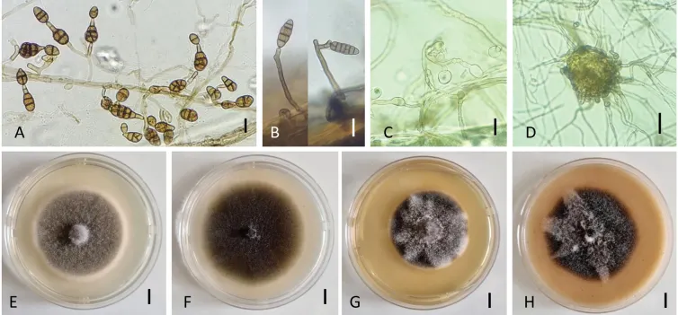
Colonies on PCA velvety to cottony colonies, mostly mycelial, subhyaline, loosely wooly, reaching 75 mm in diameter after 7 days; meager sporulation or lacking until hyphae are disturbed or scarified. Conidiophores emerging from the surface of agar or aerial vegetative hyphae scattered or clustered on cut agar in light-exposed areas. Primary conidiophores mostly simple, rarely branched, 25–75 × 5–6 µm, geniculate with 2–3(–4) conidiogenous loci. Secondary conidiophores, short, 3–7 × 3 µm, 1–2-celled, formed apically or sometimes laterally from primary conidia, mainly with one conidiogenous locus. Further geniculate extensions and conidium production yield several clusters of sporulation at the colony center. Sporulation pattern in single clumps of a few short branching chains consisting of 5–7 conidia. A high percentage of conidia solitary at any age of growth. Conidia ellipsoid to ovoid, with a rounded base and tapered apex; multiple transverse and longitudinal septa, conspicuously constricted near 2 or 3 transverse septa, in short chains. Mature conidia with 2–3 transverse septa and 0–3 longitudinal septa located mainly at the center of the colony, 20–24 × 8–15 µm; slightly older conidia, 25–34(–48) × 11–16 (–22) µm with 3–4(–5) transverse septa and 1–3 oblique or longitudinal septa. Conidial body color brown with relatively dark and thick transverse septa (Fig. 2A, B); the outer wall smooth or punctate. Formation of protoascomata and chlamydospores in the surface of vegetative mycelium conspicuous after 2–3 weeks of incubation. Chlamydospores hyaline, spherical or ovoid, terminal, sub-terminal or intercalary, measuring 13.5–22.5 × 10⁻¹⁵ µm, mostly single-celled (Fig. 2C).
Habitat dan peran ekologis
Colonies on PCA velvety to cottony colonies, mostly mycelial, subhyaline, loosely wooly, reaching 75 mm in diameter after 7 days; meager sporulation or lacking until hyphae are disturbed or scarified. Conidiophores emerging from the surface of agar or aerial vegetative hyphae scattered or clustered on cut agar in light-exposed areas. Primary conidiophores mostly simple, rarely branched, 25–75 × 5–6 µm, geniculate with 2–3(–4) conidiogenous loci. Secondary conidiophores, short, 3–7 × 3 µm, 1–2-celled, formed apically or sometimes laterally from primary conidia, mainly with one conidiogenous locus. Further geniculate extensions and conidium production yield several clusters of sporulation at the colony center. Sporulation pattern in single clumps of a few short branching chains consisting of 5–7 conidia. A high percentage of conidia solitary at any age of growth. Conidia ellipsoid to ovoid, with a rounded base and tapered apex; multiple transverse and longitudinal septa, conspicuously constricted near 2 or 3 transverse septa, in short chains. Mature conidia with 2–3 transverse septa and 0–3 longitudinal septa located mainly at the center of the colony, 20–24 × 8–15 µm; slightly older conidia, 25–34(–48) × 11–16 (–22) µm with 3–4(–5) transverse septa and 1–3 oblique or longitudinal septa.
- Habitat atau konteks temuan
- kultur yang didokumentasikan
- Substrat
- Informasi belum tersedia pada sumber rilis ini.
- Peran atau konteks
- Belum dikategorikan pada rilis ini.
Reproduksi
Colonies on PCA velvety to cottony colonies, mostly mycelial, subhyaline, loosely wooly, reaching 75 mm in diameter after 7 days; meager sporulation or lacking until hyphae are disturbed or scarified. Conidiophores emerging from the surface of agar or aerial vegetative hyphae scattered or clustered on cut agar in light-exposed areas. Primary conidiophores mostly simple, rarely branched, 25–75 × 5–6 µm, geniculate with 2–3(–4) conidiogenous loci. Secondary conidiophores, short, 3–7 × 3 µm, 1–2-celled, formed apically or sometimes laterally from primary conidia, mainly with one conidiogenous locus. Further geniculate extensions and conidium production yield several clusters of sporulation at the colony center.
Catatan identitas dan cakupan
- Nama diterima mengikuti Catalogue of Life Base2026-09-11; uraian mempertahankan konsep dan nama sumber aslinya.
- Preserve original treatment localities, hosts and substrate scope. Materials examined describe documented specimens, not the full global range.
- Impor dan terjemahan otomatis belum diperiksa editorial per profil. Struktur reproduktif yang disebutkan tidak membuktikan seluruh siklus hidup diketahui.
- Pengukuran pada uraian mengikuti spesimen, kultur, dan metode sumber; angka tidak diperluas ke semua populasi.
Catatan distribusi dalam narasi mengikuti sumbernya. Dokumentasi spesimen atau foto mengikuti catatan sumber. Bukti wilayah Indonesia dan penilaian konservasi tidak disimpulkan dari gambar.
Melihat dengan konteks
Morphology of Alternaria eureka (strain: NB968). Conidiophores and conidia on PCA for 7 days at 22 °C (A) and on inoculated carrot leaves after 24 days (B); Chlamydospores (C) and protoascomata (D) on PCA for 18 days at 22 °C. Colony phenotypes on PDA (E), PCA (F), MEA (G), and OA (H) for 7 days at 25 °C. Scale bar: 25 µm (A–D); 10 mm (E–H).
- Jenis
- Pelat ilmiah
- Kredit
- Nabahat Bessadat, Nelly Bataillé-Simoneau, Justine Colou, Bruno Hamon, Kihal Mabrouk, Philippe Simoneau
- Lisensi
- CC BY 4.0
- Sumber media
- Buka entri sumber
Sumber profil
- [1] New members of Alternaria (Pleosporales, Pleosporaceae) collected from Apiaceae in Algeria — Alternaria eureka
- [2] Catalogue of Life Base2026-09-11 — Alternaria eureka
- [3] Dokumentasi visual — Alternaria eureka
Rujukan halaman ini
Fungi. Alternaria eureka. Rilis data fungi-2026-10-12-expansion-2. https://fungi-atlas.mgadib.chatgpt.site/jamur/alternaria-eureka/.
Status: impor sumber; pemeriksaan editorial per profil belum selesai. Untuk klaim biologis, gunakan rujukan pada bagian terkait dan periksa konteks sumber aslinya.