Ciri dan struktur
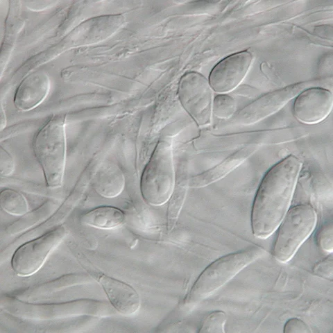
The colonies of Aphanoascus fulvescens at 30 C on glucose-peptone agar have a flat topology and a powdery to felt-like texture and are usually white to cream coloured depending on the number of brown ascomata present on the surface. Under the microscope, A. fulvescens is seen to have numerous, large club-shaped conidia with older colonies containing fruiting bodies, or ascomata, at their centres. These fruiting bodies are relatively large, ranging from 290–500 μm in diameter, and are either colourless or have a light brown pigment. The presence of the ascomata is characteristic of all members of the Ascomycota. The ascomata are smooth and have thick, enclosed walls lacking any openings. They reach full maturity after 8–10 days of colonial growth at optimal temperatures and eventually burst to release the asci contained within them. The asci have walls composed of two to four layers of flattened cells which in total amount to 4–6 μm in thickness. There are eight ascospores contained within each ascus in A. fulvescens colonies. The ellipsoidal ascospores are a light brown colour and are either lens shaped or disc shaped. They can be as large as 5 x 3.5 μm and have notably rough walls. The ascospores of A. fulvescens are different than those of other members of the Amauroascaeae which have ascospores that are usually globular and round. Rather, the unicellular ascospores of A. fulvescens are found to be flattened with reticulate walls. The hyphae of A. fulvescens are hyaline, branched, and contain many cross-walls (setpa). They are generally 1.7–3 μm wide and are contained within thin walls.
Habitat dan peran ekologis
Aphanoascus fulvescens is commonly found in the soil and in dung, living as a keratinophilic saprotroph. It is also often isolated from keratin-rich tissues such as hair and nails that have been discarded from the host. A. fulvescens is a geophilic fungus and therefore does not normally infect mammals. The organism is more commonly isolated from soil that has been inhabited frequently by animals than soil that has not. However, while this fungus is not a true dermatophyte, it is opportunistic and has been seen to cause dermatophytosis in humans and other mammals on occasion. A. fulvescens has a worldwide distribution, however it is more frequently isolated in temperate climates. This is because the organism has optimal growth at 28 °C at which it grows 3–4 mm per day and up to 25 mm per week. At elevated temperatures, such as that of the human body (37 °C), A. fulvescens shows restricted growth, averaging only 5–6 mm of growth over the course of seven days. This may be a possible explanation as to why infections involving A. fulvescens are uncommon in humans and other mammals.
- Habitat atau konteks temuan
- Informasi belum tersedia pada sumber rilis ini.
- Substrat
- Informasi belum tersedia pada sumber rilis ini.
- Peran atau konteks
- Pengurai
Reproduksi
The colonies of Aphanoascus fulvescens at 30 C on glucose-peptone agar have a flat topology and a powdery to felt-like texture and are usually white to cream coloured depending on the number of brown ascomata present on the surface. Under the microscope, A. fulvescens is seen to have numerous, large club-shaped conidia with older colonies containing fruiting bodies, or ascomata, at their centres. These fruiting bodies are relatively large, ranging from 290–500 μm in diameter, and are either colourless or have a light brown pigment. The presence of the ascomata is characteristic of all members of the Ascomycota. The ascomata are smooth and have thick, enclosed walls lacking any openings. They reach full maturity after 8–10 days of colonial growth at optimal temperatures and eventually burst to release the asci contained within them.
Catatan identitas dan cakupan
- Nama diterima mengikuti Catalogue of Life Base2026-09-11; uraian mempertahankan konsep dan nama sumber aslinya.
- Original geographic, specimen and life-cycle scope retained; source import awaits individual editorial review.
- Impor dan terjemahan otomatis belum diperiksa editorial per profil. Struktur reproduktif yang disebutkan tidak membuktikan seluruh siklus hidup diketahui.
- Pengukuran pada uraian mengikuti spesimen, kultur, dan metode sumber; angka tidak diperluas ke semua populasi.
Catatan distribusi dalam narasi mengikuti sumbernya. Dokumentasi spesimen atau foto mengikuti catatan sumber. Bukti wilayah Indonesia dan penilaian konservasi tidak disimpulkan dari gambar.
Melihat dengan konteks
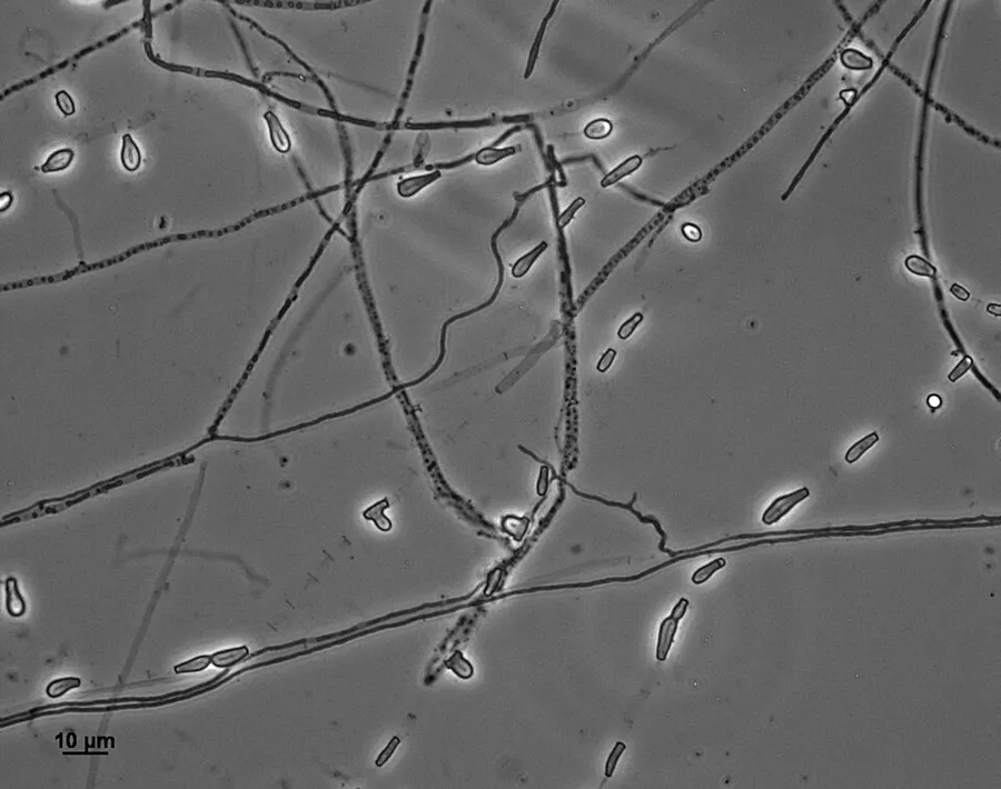
Dokumentasi Aphanoascus fulvescens dari Wikimedia Commons. Aphanoascus fulvescens (UAMH 11590) Slide culture incubated at 25°C for 41 days, photographed in phase contrast microscopy. Identifikasi mengikuti record berkas sumber.
- Jenis
- Fotografi
- Kredit
- Medmyco
- Lisensi
- CC BY-SA 4.0
- Sumber media
- Buka entri sumber
Sumber profil
- [1] Aphanoascus fulvescens — Wikipedia (en)
- [2] Catalogue of Life Base2026-09-11 — Aphanoascus fulvescens
- [3] Dokumentasi visual — Aphanoascus fulvescens
Rujukan halaman ini
Fungi. Aphanoascus fulvescens. Rilis data fungi-2026-10-12-expansion-2. https://fungi-atlas.mgadib.chatgpt.site/jamur/aphanoascus-fulvescens/.
Status: impor sumber; pemeriksaan editorial per profil belum selesai. Untuk klaim biologis, gunakan rujukan pada bagian terkait dan periksa konteks sumber aslinya.