01 / Morfologi

Ciri dan struktur

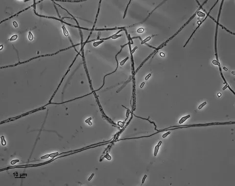
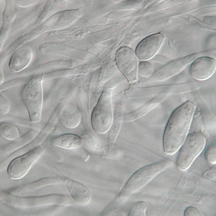
Chrysosporium keratinophilum colonies grow rapidly at 25 °C approximately 60–100 mm in 21 days. Colonies can be flat or folded, dry, powdery, or velvety with a white- or cream-coloured center The colony surface is dotted with droplets of clear or brown exuded liquid. The hyphae are septate and the conidia are hyaline, broad-based and one-celled. The conidia are large, smooth to slightly rough-walled, sometimes slightly curved and occasionally septate. The conidia are broadly "light-bulb" shaped with an abruptly flattened smooth base. Colonies grow to about 30 mm in diameter in one week and are flat with a powdery to suede-like texture. The colony reverse is also cream-coloured. Chrysosporium keratinophilum produces abundant aleurioconidia that resemble the microconidia of dermatophytes; however, the conidia of C. keratinophilum are considerably larger. Chrysosporium keratinophilum has been associated with two closely related sexual states: Aphanoascus keratinophilus and Aphanoascus fulvescens.

Sebagian uraian ditampilkan dalam bahasa sumber; terjemahannya belum tersedia atau belum lolos pemeriksaan.

Rujukan: [1]

Pengukuran mengikuti objek, spesimen atau kultur, dan metode yang disebutkan sumber. Baca uraian asli untuk konteks lengkap.

02 / Kehidupan

Habitat dan peran ekologis

Chrysosporium keratinophilum is often referred to as a keratinophilic fungus in reference to its affinity for growth on keratin-rich non-living materials such as skin scales and hairs separated from the host. Chrysosporium keratinophilum produces a keratin-degrading enzyme that functions at 90 °C. Its process of digestion occurs in two stages, requiring keratin to be chemically altered to a structureless form before it is digested. The method of hair digestion is carried out with perforating bodies. Chrysosporium spp. are asexual states of fungi in the genera Aphanoascus, Nannizziopsis, and Uncinocarpus. The fungus commonly grows on feathers, hooves, hair and other dead matter. It is rarely found on human skin and more commonly found in soil in temperate areas, plant material, dung and on birds. A study on keratinophylic fungi in the water sediments of India, by Katiyar and Kushwaha, found C. keratinophilum in sediments of catch basins and sewage sludge in India and Poland. Chrysosporium keratinophilum is associated to mud sludge structure, high humidity, volatile solids, low carbon nitrogen ratios and tolerance to heavy metals. Together, these give C. keratinophilum a high long-term survival probability in superficial water which may present an exposure risk, especially to people in India who bathe in these waters.

Sebagian uraian ditampilkan dalam bahasa sumber; terjemahannya belum tersedia atau belum lolos pemeriksaan.

Rujukan: [1]

Habitat atau konteks temuan
Informasi belum tersedia pada sumber rilis ini.
Substrat
tanah
Peran atau konteks
Belum dikategorikan pada rilis ini.
Temukan hubungan serupa
03 / Siklus hidup

Reproduksi

Chrysosporium keratinophilum colonies grow rapidly at 25 °C approximately 60–100 mm in 21 days. Colonies can be flat or folded, dry, powdery, or velvety with a white- or cream-coloured center The colony surface is dotted with droplets of clear or brown exuded liquid. The hyphae are septate and the conidia are hyaline, broad-based and one-celled. The conidia are large, smooth to slightly rough-walled, sometimes slightly curved and occasionally septate. The conidia are broadly "light-bulb" shaped with an abruptly flattened smooth base. Colonies grow to about 30 mm in diameter in one week and are flat with a powdery to suede-like texture. The colony reverse is also cream-coloured. Chrysosporium keratinophilum produces abundant aleurioconidia that resemble the microconidia of dermatophytes; however, the conidia of C. keratinophilum are considerably larger.

Sebagian uraian ditampilkan dalam bahasa sumber; terjemahannya belum tersedia atau belum lolos pemeriksaan.

Rujukan: [1]

Sinonim menurut acuan taksonomi rilis: Chrysosporium keratinophilum
04 / Batas informasi

Catatan identitas dan cakupan

  • Nama diterima mengikuti Catalogue of Life Base2026-09-11; uraian mempertahankan konsep dan nama sumber aslinya.
  • Original geographic, specimen and life-cycle scope retained; source import awaits individual editorial review.
  • Impor dan terjemahan otomatis belum diperiksa editorial per profil. Struktur reproduktif yang disebutkan tidak membuktikan seluruh siklus hidup diketahui.
  • Pengukuran pada uraian mengikuti spesimen, kultur, dan metode sumber; angka tidak diperluas ke semua populasi.

Catatan distribusi dalam narasi mengikuti sumbernya. Dokumentasi spesimen atau foto mengikuti catatan sumber. Bukti wilayah Indonesia dan penilaian konservasi tidak disimpulkan dari gambar.

05 / Dokumentasi

Melihat dengan konteks

Mikrograf Aphanoascus keratinophilus yang dicatat sumber sebagai Chrysosporium keratinophilum, sinonim dalam profil ini. Foto: J Scott / Medmyco.

Jenis
Mikrograf
Kredit
J Scott (foto pribadi); Medmyco at English Wikipedia
Lisensi
CC0 1.0
Sumber media
Buka entri sumber

Gambar menunjukkan objek menurut identifikasi sumber, bukan verifikasi independen spesimen oleh Fungi. Tidak ada skala baru yang ditambahkan.

06 / Rujukan

Sumber profil

  1. [1] Chrysosporium keratinophilum — Wikipedia (en)Bagian sumber: Frozen revision 1373213246
    Tanggal penghimpunan sumber: 2026-10-12
    Teks sumber: CC BY-SA4.0 · Wikipedia contributors
  2. [2] Catalogue of Life Base2026-09-11 — Aphanoascus keratinophilusBagian sumber: ColDP NameUsage.tsv ID FB7N; accepted species; source 2073
    Tanggal penghimpunan sumber: 2026-10-12
  3. [3] Dokumentasi visual — Aphanoascus keratinophilusBagian sumber: Identitas mengikuti sumber dokumentasi; bukan penentuan spesimen independen.
    Tanggal penghimpunan sumber: 2026-10-17
    Media sumber: CC0 1.0 · J Scott (foto pribadi); Medmyco at English Wikipedia

Adaptasi teks: CC BY-SA 4.0. Wikipedia contributors — Chrysosporium keratinophilum — Wikipedia (en)

Uraian asli dan riwayat sumber

Riwayat atau halaman sumber

Ringkasan sumber

Chrysosporium keratinophilum is a mold that is closely related to the dermatophytic fungi (family Arthrodermataceae) and is mainly found in soil and the coats of wild animals to break down keratin. Chrysosporium keratinophilum is one of the more commonly occurring species of the genus Chrysosporium in nature. It is easily detected due to its characteristic "light-bulb" shape and flat base. Chrysosporium keratinophilum is most commonly found in keratin-rich, dead materials such as feathers, skin scales, hair, and hooves.

Revisi sumber

Morfologi sumber

Chrysosporium keratinophilum colonies grow rapidly at 25 °C approximately 60–100 mm in 21 days. Colonies can be flat or folded, dry, powdery, or velvety with a white- or cream-coloured center The colony surface is dotted with droplets of clear or brown exuded liquid. The hyphae are septate and the conidia are hyaline, broad-based and one-celled. The conidia are large, smooth to slightly rough-walled, sometimes slightly curved and occasionally septate. The conidia are broadly "light-bulb" shaped with an abruptly flattened smooth base. Colonies grow to about 30 mm in diameter in one week and are flat with a powdery to suede-like texture. The colony reverse is also cream-coloured. Chrysosporium keratinophilum produces abundant aleurioconidia that resemble the microconidia of dermatophytes; however, the conidia of C. keratinophilum are considerably larger. Chrysosporium keratinophilum has been associated with two closely related sexual states: Aphanoascus keratinophilus and Aphanoascus fulvescens.

Revisi sumber

Ekologi sumber

Chrysosporium keratinophilum is often referred to as a keratinophilic fungus in reference to its affinity for growth on keratin-rich non-living materials such as skin scales and hairs separated from the host. Chrysosporium keratinophilum produces a keratin-degrading enzyme that functions at 90 °C. Its process of digestion occurs in two stages, requiring keratin to be chemically altered to a structureless form before it is digested. The method of hair digestion is carried out with perforating bodies. Chrysosporium spp. are asexual states of fungi in the genera Aphanoascus, Nannizziopsis, and Uncinocarpus. The fungus commonly grows on feathers, hooves, hair and other dead matter. It is rarely found on human skin and more commonly found in soil in temperate areas, plant material, dung and on birds. A study on keratinophylic fungi in the water sediments of India, by Katiyar and Kushwaha, found C. keratinophilum in sediments of catch basins and sewage sludge in India and Poland. Chrysosporium keratinophilum is associated to mud sludge structure, high humidity, volatile solids, low carbon nitrogen ratios and tolerance to heavy metals. Together, these give C. keratinophilum a high long-term survival probability in superficial water which may present an exposure risk, especially to people in India who bathe in these waters.

Revisi sumber

Struktur reproduksi sumber

Chrysosporium keratinophilum colonies grow rapidly at 25 °C approximately 60–100 mm in 21 days. Colonies can be flat or folded, dry, powdery, or velvety with a white- or cream-coloured center The colony surface is dotted with droplets of clear or brown exuded liquid. The hyphae are septate and the conidia are hyaline, broad-based and one-celled. The conidia are large, smooth to slightly rough-walled, sometimes slightly curved and occasionally septate. The conidia are broadly "light-bulb" shaped with an abruptly flattened smooth base. Colonies grow to about 30 mm in diameter in one week and are flat with a powdery to suede-like texture. The colony reverse is also cream-coloured. Chrysosporium keratinophilum produces abundant aleurioconidia that resemble the microconidia of dermatophytes; however, the conidia of C. keratinophilum are considerably larger.

Revisi sumber

Rujukan halaman ini

Fungi. Aphanoascus keratinophilus. Rilis data fungi-2026-10-12-expansion-2. https://fungi-atlas.mgadib.chatgpt.site/jamur/aphanoascus-keratinophilus/.

Status: impor sumber; pemeriksaan editorial per profil belum selesai. Untuk klaim biologis, gunakan rujukan pada bagian terkait dan periksa konteks sumber aslinya.