Ciri dan struktur
Distinguished from other Apophysomyces species by the slightly trapezoidal sporangiospores, and from A. elegans, A. trapeziformis, and A. mexicanus by its narrower sporangiospores. Colonies on PDA attaining a diameter of 90 mm after 2 d at 37 °C, whitish at first, becoming white to cream-colored, reverse concolorous (Fig. 4A). Colonies on MEA attaining a diameter of 90 mm after 5 d at 37 °C, flat, whitish, reverse concolorous (Fig. 4B). Colonies on CZA attaining a diameter of 90 mm after 4 d at 37 °C, whitish at first, becoming white to cream-colored, with scarce aerial mycelium, reverse concolorous (Fig. 4C). On all agar media the hyphae are branched, hyaline, smooth-walled, and have 5–15 µm in diameter (Fig. 4D). Sporulation was observed only on CZA. Sporangiophores erect, usually arising singly, emerging from aerial hyphae, at first hyaline but soon becoming light brown, usually straight, slightly tapered towards the apex, unbranched, 60–890 µm in length, 3.75–7.5 µm wide, and smooth-walled. Sporangia apophysate, terminal, pyriform, multispored, white at first, becoming light greyish brown when mature, and 25–58 µm in diameter. Apophyses short, funnel to bell shaped, 21–52 × 19–46 µm (Fig. 4E). Sporangiospores slightly trapezoidal in side view, cylindrical in front view, with flattened to slightly concave lateral walls, hyaline to light brown in mass, smooth- and thin-walled, 5–6(9) × 2–3 µm (Fig. 4F).
Habitat dan peran ekologis
THAILAND. Chiang Mai Province: Mae Wang District, (18°36'46"N, 98°46'30"E), isolated from soil in agricultural area, 8 August 2017, S. Khuna, dried cultures: SDBR-CMUS26; ex-type living culture: TBRC9299 Distinguished from other Apophysomyces species by the slightly trapezoidal sporangiospores, and from A. elegans, A. trapeziformis, and A. mexicanus by its narrower sporangiospores. Colonies on PDA attaining a diameter of 90 mm after 2 d at 37 °C, whitish at first, becoming white to cream-colored, reverse concolorous (Fig. 4A). Colonies on MEA attaining a diameter of 90 mm after 5 d at 37 °C, flat, whitish, reverse concolorous (Fig. 4B). Colonies on CZA attaining a diameter of 90 mm after 4 d at 37 °C, whitish at first, becoming white to cream-colored, with scarce aerial mycelium, reverse concolorous (Fig. 4C). On all agar media the hyphae are branched, hyaline, smooth-walled, and have 5–15 µm in diameter (Fig. 4D). Sporulation was observed only on CZA. Sporangiophores erect, usually arising singly, emerging from aerial hyphae, at first hyaline but soon becoming light brown, usually straight, slightly tapered towards the apex, unbranched, 60–890 µm in length, 3.75–7.5 µm wide, and smooth-walled.
- Habitat atau konteks temuan
- kultur yang didokumentasikan
- Substrat
- Informasi belum tersedia pada sumber rilis ini.
- Peran atau konteks
- Belum dikategorikan pada rilis ini.
Reproduksi
Distinguished from other Apophysomyces species by the slightly trapezoidal sporangiospores, and from A. elegans, A. trapeziformis, and A. mexicanus by its narrower sporangiospores. Colonies on PDA attaining a diameter of 90 mm after 2 d at 37 °C, whitish at first, becoming white to cream-colored, reverse concolorous (Fig. 4A). Colonies on MEA attaining a diameter of 90 mm after 5 d at 37 °C, flat, whitish, reverse concolorous (Fig. 4B). Colonies on CZA attaining a diameter of 90 mm after 4 d at 37 °C, whitish at first, becoming white to cream-colored, with scarce aerial mycelium, reverse concolorous (Fig. 4C). On all agar media the hyphae are branched, hyaline, smooth-walled, and have 5–15 µm in diameter (Fig. 4D).
Catatan identitas dan cakupan
- Nama diterima mengikuti Catalogue of Life Base2026-09-11; uraian mempertahankan konsep dan nama sumber aslinya.
- Preserve original treatment localities, hosts and substrate scope. Materials examined describe documented specimens, not the full global range.
- Impor dan terjemahan otomatis belum diperiksa editorial per profil. Struktur reproduktif yang disebutkan tidak membuktikan seluruh siklus hidup diketahui.
- Pengukuran pada uraian mengikuti spesimen, kultur, dan metode sumber; angka tidak diperluas ke semua populasi.
Catatan distribusi dalam narasi mengikuti sumbernya. Dokumentasi spesimen atau foto mengikuti catatan sumber. Bukti wilayah Indonesia dan penilaian konservasi tidak disimpulkan dari gambar.
Melihat dengan konteks
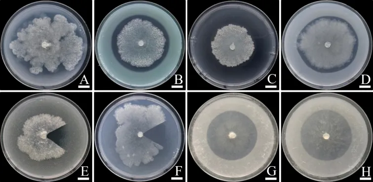
Solubilization of non-soluble minerals in agar media by Apophysomyces thailandensis SDBR-CMUS26 (holotype). A Ca 3 (PO 4) 2 B CuCO 3 •Cu(OH) 2 C CuO D ZnCO 3 E FePO 4 F MnO G Feldspar H Kaolin. Scale bars: 10 mm. Fungal colonies in E and F were cut for the solubilization area (halo zone) observation.
- Jenis
- Pelat ilmiah
- Kredit
- Surapong Khuna, Nakarin Suwannarach, Jaturong Kumla, Jomkhwan Meerak, Wipornpan Nuangmek, Tanongkiat Kiatsiriroat, Saisamorn Lumyong
- Lisensi
- CC BY 4.0
- Sumber media
- Buka entri sumber
Dokumentasi tambahan
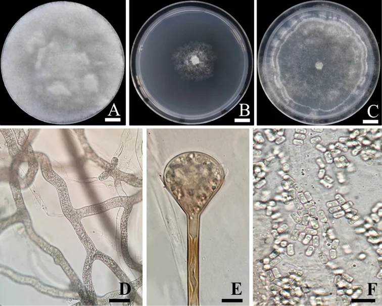
Apophysomyces thailandensis SDBR-CMUS26 (holotype). A colony on potato dextrose agar B Colony on malt extract agar C Colony on Czapek agar D Branched, aseptate hyphae E Funnel-shaped apophysis F Slightly trapezoidal sporangiospores. Scale bars: 10 mm (A–C), 10 µm (D–E), 20 µm (F).
Kredit dan penggunaan
Surapong Khuna, Nakarin Suwannarach, Jaturong Kumla, Jomkhwan Meerak, Wipornpan Nuangmek, Tanongkiat Kiatsiriroat, Saisamorn Lumyong
Ukuran dan format gambar disesuaikan tanpa pemotongan, retus, atau penambahan skala. Lisensi sumber dipertahankan.
Sumber profil
- [1] Apophysomyces thailandensis (Mucorales, Mucoromycota), a new species isolated from soil in northern Thailand and its solubilization of non-soluble minerals — Apophysomyces thailandensis
- [2] Catalogue of Life Base2026-09-11 — Apophysomyces thailandensis
- [3] Dokumentasi visual — Apophysomyces thailandensis
- [4] Dokumentasi visual — Apophysomyces thailandensis
Rujukan halaman ini
Fungi. Apophysomyces thailandensis. Rilis data fungi-2026-10-12-expansion-2. https://fungi-atlas.mgadib.chatgpt.site/jamur/apophysomyces-thailandensis/.
Status: impor sumber; pemeriksaan editorial per profil belum selesai. Untuk klaim biologis, gunakan rujukan pada bagian terkait dan periksa konteks sumber aslinya.