Ciri dan struktur
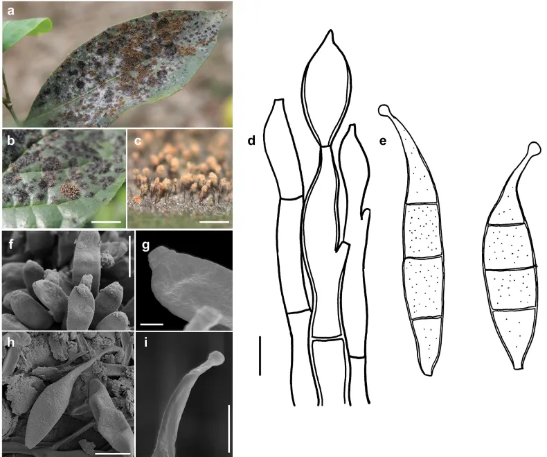
Colonies effuse, rust brown or pale brown, with hyphae that form large, erect, dark synnemata clearly visible under the stereomicroscope, but sometimes only loose unstalked tufts around the tips of the setae of the meliolalean host. Hyphae superficial, branched, septate, thin-walled, 1–2.5 µm wide, smooth. Conidiophores may form straw-coloured or pale olivaceous synnemata up to 1.5 mm long, 40 µm wide at the basal stalk-like part. Sometimes the synnemata grow around and up the setae of the meliolalean host. Individual conidiophores straight or sometimes flexuous, cylindrical, 2.5–5 µm thick towards the apex, pale olivaceous brown, with denticles. Conidia solitary, straight or slightly curved, fusiform, truncate at the base, tapering towards the apex and often terminating in a little bulbous swelling, 1 to mostly 3 septate, thin-walled, variable in size, (17–)30–37(–80) × (3.5–)7–8.5 µm, at first more or less colourless, at maturity becoming pale straw coloured, minutely rough-walled. As seen by SEM, the ornamentation of the surface of the conidia is distinctly reticulated, with thin networks and no ridges.
Habitat dan peran ekologis
On colonies of Amazonia spp., Asteridiella spp., Irenopsis spp. and Meliola spp. on living leaves of various plants in Congo, Ghana, Guinea, India, Mauritius, Nigeria, Perú, Philippines, Puerto Rico, Sierra Leone, St. Thomé, Taiwan, Tanzania, Uganda, U.S.A. and Venezuela. Only one single collection on Balladyna sp. (Balladynaceae, Dothideomycetes) as a fungal host (Deighton and Pirozynski 1972). Atractilina parasitica is reported here for the first time for Benin.
- Habitat atau konteks temuan
- Informasi belum tersedia pada sumber rilis ini.
- Substrat
- Informasi belum tersedia pada sumber rilis ini.
- Peran atau konteks
- Belum dikategorikan pada rilis ini.
Reproduksi
Colonies effuse, rust brown or pale brown, with hyphae that form large, erect, dark synnemata clearly visible under the stereomicroscope, but sometimes only loose unstalked tufts around the tips of the setae of the meliolalean host. Hyphae superficial, branched, septate, thin-walled, 1–2.5 µm wide, smooth. Conidiophores may form straw-coloured or pale olivaceous synnemata up to 1.5 mm long, 40 µm wide at the basal stalk-like part. Sometimes the synnemata grow around and up the setae of the meliolalean host. Individual conidiophores straight or sometimes flexuous, cylindrical, 2.5–5 µm thick towards the apex, pale olivaceous brown, with denticles. Conidia solitary, straight or slightly curved, fusiform, truncate at the base, tapering towards the apex and often terminating in a little bulbous swelling, 1 to mostly 3 septate, thin-walled, variable in size, (17–)30–37(–80) × (3.5–)7–8.5 µm, at first more or less colourless, at maturity becoming pale straw coloured, minutely rough-walled.
Catatan identitas dan cakupan
- Nama diterima mengikuti Catalogue of Life Base2026-09-11; uraian mempertahankan konsep dan nama sumber aslinya.
- Preserve original treatment localities, hosts and substrate scope. Materials examined describe documented specimens, not the full global range.
- Impor dan terjemahan otomatis belum diperiksa editorial per profil. Struktur reproduktif yang disebutkan tidak membuktikan seluruh siklus hidup diketahui.
- Pengukuran pada uraian mengikuti spesimen, kultur, dan metode sumber; angka tidak diperluas ke semua populasi.
Catatan distribusi dalam narasi mengikuti sumbernya. Dokumentasi spesimen atau foto mengikuti catatan sumber. Bukti wilayah Indonesia dan penilaian konservasi tidak disimpulkan dari gambar.
Melihat dengan konteks
Atractilina parasitica (MB127, MB136) a synnemata (gold spots) on colonies of Meliola sp. (black spots) on a leaf of Opilia celtidifolia b synnemata of (gold spots) on colonies of Meliola clerodendricola (black spots) on a leaf of Clerodendrum capitatum c synnemata d conidiophores drawn in optical section. The thickness of the wall is indicated only in the drawing in the middle e conidia shown in optical section f–i as seen by SEM f conidiophores with denticles g a denticle at the tip of a conidiophore h conidium i bulbous swelling at the tip of a conidium. Scale bars: 1.5 mm (b); 1 mm (c); 5 μm (d,e,i); 8 μm (f); 1 μm (g); 6 μm (h).
- Jenis
- Pelat ilmiah
- Kredit
- Miguel A. Bermúdez-Cova, Tina A. Hofmann, Nourou S. Yorou, Meike Piepenbring
- Lisensi
- CC BY 4.0
- Sumber media
- Buka entri sumber
Sumber profil
- [1] Systematic revision of species of Atractilina and Spiropes hyperparasitic on Meliolales (Ascomycota) in the tropics — Atractilina parasitica
- [2] Catalogue of Life Base2026-09-11 — Atractilina parasitica
- [3] Dokumentasi visual — Atractilina parasitica
Rujukan halaman ini
Fungi. Atractilina parasitica. Rilis data fungi-2026-10-12-expansion-2. https://fungi-atlas.mgadib.chatgpt.site/jamur/atractilina-parasitica/.
Status: impor sumber; pemeriksaan editorial per profil belum selesai. Untuk klaim biologis, gunakan rujukan pada bagian terkait dan periksa konteks sumber aslinya.