Ciri dan struktur
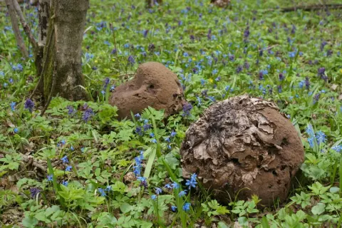
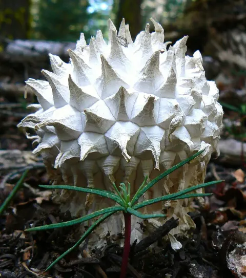
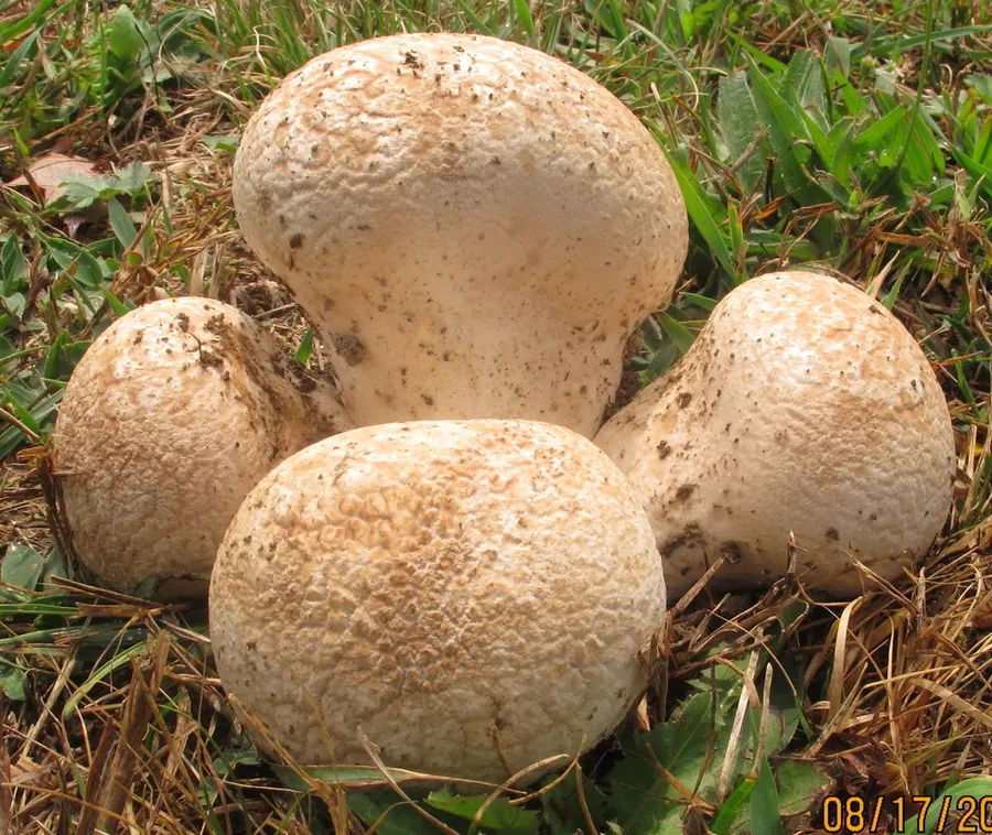
The fruit bodies of Calvatia craniiformis grow to dimensions of 6 – 20 cm tall by 8 – 20 cm wide, and have a form ranging from pear-shaped, to flattened-spherical, to obovate (roughly egg-shaped), to tunicate (like an inverted cone). At the bottom is a thick, often crumpled base attached to a cord-like rhizomorph, which is often encrusted with surrounding soil. The rhizomorphs are well developed, and when cut into longitudinal section, reveal three distinct tissues: an outer cortex, a subcortical layer, and a central core. The thin and fragile exoperidium (the outer layer of "skin") is whitish-gray to gray, and initially smooth before becoming areolate (divided by cracks into discrete areas). The base extends up one-third to one-halfway into the puffball (where it becomes the columnella) tapering to a point. The gleba is initially whitish, and then yellow-green, and finally brownish-green in older specimens with mature spores. Spores are spherical, hyaline (translucent), and measure 2.5–3.4 μm in diameter. They are thick-walled with a short pedicel (a tubelike extension), and are ornamented with tiny spines (verrucae) that are roughly equidistant from each other. Capillitial threads are long, hyaline, and branched, measuring 2.4–4 μm thick. They are septate and occasionally have pits on their walls. The exoperidium comprises thick-walled, inflated hyphae mixed with sphaerocysts (spherical cells), while the endoperidium is made of tightly interwoven, thick-walled hyphae. In the rhizomorphs, the hyphae in the central core are several times as thick as those in the surrounding subcortex.
Habitat dan peran ekologis
Calvatia craniiformis, commonly known as the brain puffball or the skull-shaped puffball, is a species of puffball fungus in the family Agaricaceae. It is found in Asia, Australia, and North America, where it grows on the ground in open woods. Its name, derived from the same Latin root as cranium, alludes to its resemblance to an animal's brain. The skull-shaped fruit body is 8 – 20 cm broad by 6 – 20 cm tall and white to tan. Initially smooth, the skin (peridium) develops wrinkles and folds as it matures, cracking and flaking with age. The peridium eventually sloughs away, exposing a powdery yellow-brown to greenish-yellow spore mass (the gleba). The puffball is edible when the gleba is still white and firm, before it matures to become yellow-brown and powdery. Mature specimens have been used in the traditional or folk medicines of China, Japan, and the Ojibwe as a hemostatic or wound dressing agent. Several bioactive compounds have been isolated and identified from the brain puffball. Fruit bodies arise from the tips or the lateral branches or rhizomorphs. New fruit bodies are made of an exterior of thick hyphae similar to those found in the rhizomorph core; in contrast, the hyphae of the interior originate from the puffball's core.
- Habitat atau konteks temuan
- Informasi belum tersedia pada sumber rilis ini.
- Substrat
- tanah
- Peran atau konteks
- Belum dikategorikan pada rilis ini.
Reproduksi
The fruit bodies of Calvatia craniiformis grow to dimensions of 6 – 20 cm tall by 8 – 20 cm wide, and have a form ranging from pear-shaped, to flattened-spherical, to obovate (roughly egg-shaped), to tunicate (like an inverted cone). At the bottom is a thick, often crumpled base attached to a cord-like rhizomorph, which is often encrusted with surrounding soil. The rhizomorphs are well developed, and when cut into longitudinal section, reveal three distinct tissues: an outer cortex, a subcortical layer, and a central core. The thin and fragile exoperidium (the outer layer of "skin") is whitish-gray to gray, and initially smooth before becoming areolate (divided by cracks into discrete areas).
Catatan identitas dan cakupan
- Nama diterima mengikuti Catalogue of Life Base2026-09-11; uraian mempertahankan konsep dan nama sumber aslinya.
- Original geographic, specimen and life-cycle scope retained; source import awaits individual editorial review.
- Impor dan terjemahan otomatis belum diperiksa editorial per profil. Struktur reproduktif yang disebutkan tidak membuktikan seluruh siklus hidup diketahui.
- Pengukuran pada uraian mengikuti spesimen, kultur, dan metode sumber; angka tidak diperluas ke semua populasi.
Catatan distribusi dalam narasi mengikuti sumbernya. Dokumentasi spesimen atau foto mengikuti catatan sumber. Bukti wilayah Indonesia dan penilaian konservasi tidak disimpulkan dari gambar.
Melihat dengan konteks
Dokumentasi Calvatia craniiformis menurut asosiasi foto pada record takson iNaturalist. Kredit: mycowalt. Identitas mengikuti sumber; bukan penentuan spesimen independen.
- Jenis
- Fotografi
- Kredit
- (c) mycowalt, some rights reserved (CC BY-SA), uploaded by mycowalt
- Lisensi
- CC BY-SA 4.0
- Sumber media
- Buka entri sumber
Dokumentasi tambahan
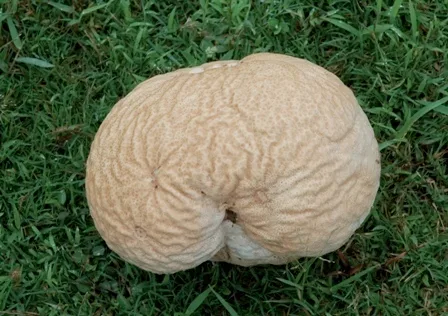
Foto yang diasosiasikan dengan Calvatia craniiformis pada iNaturalist. Identifikasi mengikuti sumber; bukan penentuan spesimen independen.
Kredit dan penggunaan
(c) Sindhu Ramchandran, the mushroom was identified by Mr Marco Della Maggiora, Italy, some rights reserved (CC BY-SA)
Ukuran dan format gambar disesuaikan tanpa pemotongan, retus, atau penambahan skala. Lisensi sumber dipertahankan.
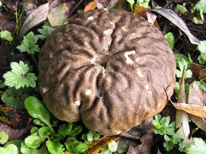
Foto yang diasosiasikan dengan Calvatia craniiformis pada iNaturalist. Identifikasi mengikuti sumber; bukan penentuan spesimen independen.
Kredit dan penggunaan
(c) David Whyte, some rights reserved (CC BY-SA), uploaded by David Whyte
Ukuran dan format gambar disesuaikan tanpa pemotongan, retus, atau penambahan skala. Lisensi sumber dipertahankan.
Sumber profil
- [1] Calvatia craniiformis — Wikipedia (en)
- [2] Catalogue of Life Base2026-09-11 — Calvatia craniiformis
- [3] Dokumentasi visual — Calvatia craniiformis
- [4] Dokumentasi visual — Calvatia craniiformis
- [5] Dokumentasi visual — Calvatia craniiformis
Rujukan halaman ini
Fungi. Calvatia craniiformis. Rilis data fungi-2026-10-12-expansion-2. https://fungi-atlas.mgadib.chatgpt.site/jamur/calvatia-craniiformis/.
Status: impor sumber; pemeriksaan editorial per profil belum selesai. Untuk klaim biologis, gunakan rujukan pada bagian terkait dan periksa konteks sumber aslinya.