Ciri dan struktur
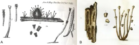
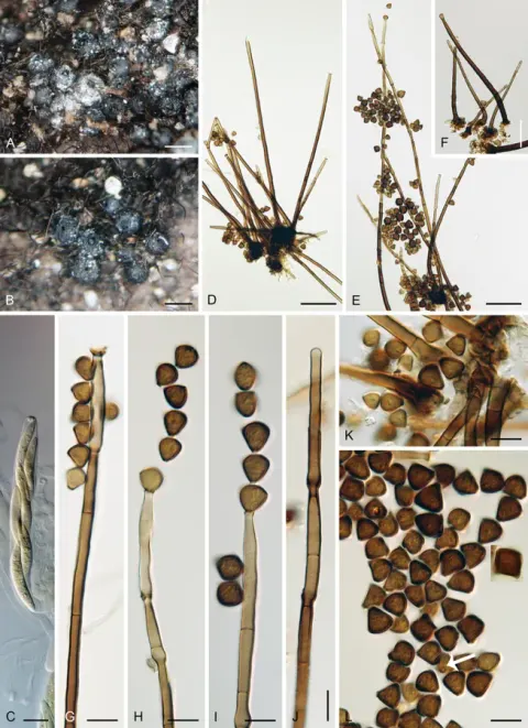
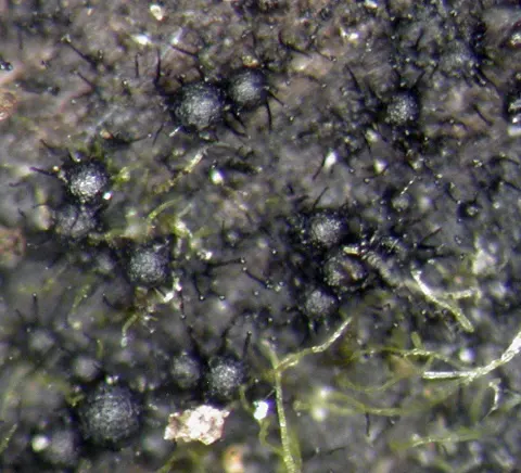
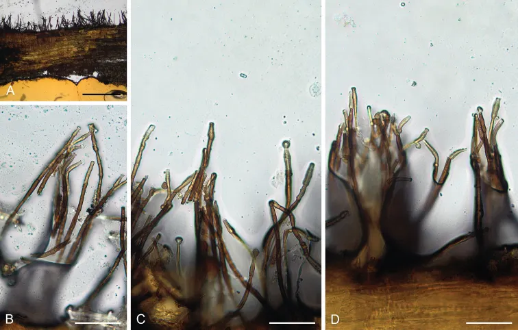
Colonies on natural substrate effuse, hairy to velutinous, dark brown to almost black. Anamorph. Conidiophores 77–220 × 4.5–6(–7) μm wide, 7–8 μm above the base, macronematous, solitary or arise in tufts, erect, straight or slightly flexuous, unbranched, dark brown, paler towards the apex, septate. Capitate hyphae absent. Conidiogenous cells 18–25 × 3.5–4.5 μm tapering to ca. 2.5 μm, integrated, terminal, monophialidic, extending percurrently, obclavate to subcylindrical or slightly lageniform, pale brown, paler towards the apex; collarettes 3–4 μm wide, 1.5(–2) μm deep, funnel-shaped, subhyaline, smooth, margin entire. Conidia 6–8(–9) μm long, 4.5–6(–7) μm wide at the apical end, ca. 2 μm wide at the basal hilum (mean ± SD = 7.4 ± 1.1 × 6.0 ± 1.2 μm × 2.0 ± 0.0 μm), rounded-obconic in side view, with three blunt corners when viewed from above, broadly rounded to flattened at the apex, truncate at the basal scar, pale brown to pale fuscous, thick-walled, smooth; formed singly, adhered in basipetal chains. Teleomorph. Unknown.
Habitat dan peran ekologis
Saprobe on dead culms of Bambusa sp., decaying wood of Fagus sp. and other unknown hosts in freshwater and terrestrial habitats. It is known in China, India and the USA (Linder 1933; Sharma 1980; Luo et al. 2019 as C. cubensis).
- Habitat atau konteks temuan
- Informasi belum tersedia pada sumber rilis ini.
- Substrat
- Informasi belum tersedia pada sumber rilis ini.
- Peran atau konteks
- Belum dikategorikan pada rilis ini.
Reproduksi
Colonies on natural substrate effuse, hairy to velutinous, dark brown to almost black. Anamorph. Conidiophores 77–220 × 4.5–6(–7) μm wide, 7–8 μm above the base, macronematous, solitary or arise in tufts, erect, straight or slightly flexuous, unbranched, dark brown, paler towards the apex, septate. Capitate hyphae absent. Conidiogenous cells 18–25 × 3.5–4.5 μm tapering to ca. 2.5 μm, integrated, terminal, monophialidic, extending percurrently, obclavate to subcylindrical or slightly lageniform, pale brown, paler towards the apex; collarettes 3–4 μm wide, 1.5(–2) μm deep, funnel-shaped, subhyaline, smooth, margin entire. Conidia 6–8(–9) μm long, 4.5–6(–7) μm wide at the apical end, ca. 2 μm wide at the basal hilum (mean ± SD = 7.4 ± 1.1 × 6.0 ± 1.2 μm × 2.0 ± 0.0 μm), rounded-obconic in side view, with three blunt corners when viewed from above, broadly rounded to flattened at the apex, truncate at the basal scar, pale brown to pale fuscous, thick-walled, smooth; formed singly, adhered in basipetal chains.
Catatan identitas dan cakupan
- Nama diterima mengikuti Catalogue of Life Base2026-09-11; uraian mempertahankan konsep dan nama sumber aslinya.
- Preserve original treatment localities, hosts and substrate scope. Materials examined describe documented specimens, not the full global range.
- Impor dan terjemahan otomatis belum diperiksa editorial per profil. Struktur reproduktif yang disebutkan tidak membuktikan seluruh siklus hidup diketahui.
- Pengukuran pada uraian mengikuti spesimen, kultur, dan metode sumber; angka tidak diperluas ke semua populasi.
Catatan distribusi dalam narasi mengikuti sumbernya. Dokumentasi spesimen atau foto mengikuti catatan sumber. Bukti wilayah Indonesia dan penilaian konservasi tidak disimpulkan dari gambar.
Melihat dengan konteks
Catenularia angulospora (holotype FH 00965375) A vertical section of the wood with colony B–D conidiophores with conidia. Scale bars: 500 μm (A); 50 μm (B–D).
- Jenis
- Pelat ilmiah
- Kredit
- Martina Réblová, Jana Nekvindová, Andrew N. Miller
- Lisensi
- CC BY 4.0
- Sumber media
- Buka entri sumber
Sumber profil
- [1] Phylogeny and taxonomy of Catenularia and similar fungi with catenate conidia — Catenularia angulospora
- [2] Catalogue of Life Base2026-09-11 — Catenularia angulospora
- [3] Dokumentasi visual — Catenularia angulospora
Rujukan halaman ini
Fungi. Catenularia angulospora. Rilis data fungi-2026-10-12-expansion-2. https://fungi-atlas.mgadib.chatgpt.site/jamur/catenularia-angulospora/.
Status: impor sumber; pemeriksaan editorial per profil belum selesai. Untuk klaim biologis, gunakan rujukan pada bagian terkait dan periksa konteks sumber aslinya.