01 / Morfologi

Ciri dan struktur

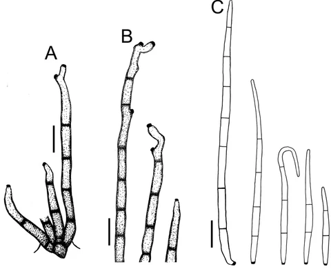
Cercospora melongenae is found primarily in warm climates close to the equator. In the US it is prevalent in Hawaii, and could potentially be found in the southern Continental US. Symptoms begin to show on the underside of older, lower leaves first due to the proximity to the soil. They are the first leaves to receive the inoculum. Lesions can be found on leaves, stems and petioles. The lesions appear small and circular at first and later progress to angular and more irregular shapes. The lesions are usually 4-10mm in diameter, and have light to dark tan concentric spots. Spots appear on the upper leaf surface first and eventually progress to the lower side of the leaf. Upon observation in the field with a handheld scope, signs of conidiophores can be seen in the stomata of infected tissue. Conidiophores are typically mid-brown, and straight or slightly flexuous and septate. They are typically 30-150 microns long and 4-7 microns wide. Conidia can also sometimes be seen. Conidia of Cercospora melongenae typically have a truncate base with a somewhat thickened convex scar and a sub-acute apex. The conidia are typically 40-150 microns long and 3-4.5 microns wide, and of 4-14 septate. Later in the disease cycle excessive sporulation can be readily observed in the center of the lesions. Eventually the lesions will dry up and in some cases fall out. The fungi does not affect the fruit, however yield can be impacted due to loss of photosynthetic material. Cercospora melongenae exists wholly in its asexual stage; even when grown in culture in the lab, no sexual stage is seen. This means that this disease has an imperfect life cycle, making it of the deuteromycota.

Sebagian uraian ditampilkan dalam bahasa sumber; terjemahannya belum tersedia atau belum lolos pemeriksaan.

Rujukan: [1]

Pengukuran mengikuti objek, spesimen atau kultur, dan metode yang disebutkan sumber. Baca uraian asli untuk konteks lengkap.

02 / Kehidupan

Habitat dan peran ekologis

Cercospora melongenae adalah patogen tumbuhan jamur yang menyebabkan noda daun pada terong (Solanum melongenum). Ini adalah jamur deuteromycete yang terutama terbatas pada spesies terong. Some other spesies inang adalah Solanum aethiopicum dan Solanum incanum. This plant pathogen only attacks leaves of eggplants and not the fruit. Ini cukup umum di antara jamur yang menginfeksi taman komunitas dan kebun rumah terong. Generally speaking, Cercospora melongenae menyerang semua varietas lokal teropong, tetapi paling parah pada Philippine eggplant dan kurang parasit pada Siamese variety. Cercospora melongenae terutama ditemukan di iklim hangat dekat khatulistiwa. Di AS, ini sangat umum di Hawaii, dan berpotensi ditemukan di Amerika Serikat Benua Selatan. Symptoms begin untuk muncul di bagian bawah daun yang lebih tua, lebih rendah pertama karena kedekatan dengan tanah. They are daun pertama untuk menerima inoculum. Lesions can ditemukan pada daun, batang dan petioles. Lesi tampak kecil dan bulat pada awalnya dan kemudian berkembang menjadi bentuk sudut dan lebih tidak teratur. Lesi biasanya berdiameter 4-10mm, dan memiliki titik konsentris berwarna cokelat terang hingga gelap. Spots appear pada permukaan atas daun pertama dan akhirnya maju ke sisi bawah daun.

Sebagian uraian ditampilkan dalam bahasa sumber; terjemahannya belum tersedia atau belum lolos pemeriksaan.

Rujukan: [1]

Habitat atau konteks temuan
Informasi belum tersedia pada sumber rilis ini.
Substrat
Informasi belum tersedia pada sumber rilis ini.
Peran atau konteks
Belum dikategorikan pada rilis ini.
Temukan hubungan serupa
03 / Siklus hidup

Reproduksi

Cercospora melongenae is found primarily in warm climates close to the equator. In the US it is prevalent in Hawaii, and could potentially be found in the southern Continental US. Symptoms begin to show on the underside of older, lower leaves first due to the proximity to the soil. They are the first leaves to receive the inoculum. Lesions can be found on leaves, stems and petioles. The lesions appear small and circular at first and later progress to angular and more irregular shapes. The lesions are usually 4-10mm in diameter, and have light to dark tan concentric spots. Spots appear on the upper leaf surface first and eventually progress to the lower side of the leaf. Upon observation in the field with a handheld scope, signs of conidiophores can be seen in the stomata of infected tissue.

Sebagian uraian ditampilkan dalam bahasa sumber; terjemahannya belum tersedia atau belum lolos pemeriksaan.

Rujukan: [1]

Sinonim menurut acuan taksonomi rilis: Cercosporina melongenae
04 / Batas informasi

Catatan identitas dan cakupan

  • Nama diterima mengikuti Catalogue of Life Base2026-09-11; uraian mempertahankan konsep dan nama sumber aslinya.
  • Original geographic, specimen and life-cycle scope retained; source import awaits individual editorial review.
  • Impor dan terjemahan otomatis belum diperiksa editorial per profil. Struktur reproduktif yang disebutkan tidak membuktikan seluruh siklus hidup diketahui.
  • Pengukuran pada uraian mengikuti spesimen, kultur, dan metode sumber; angka tidak diperluas ke semua populasi.

Catatan distribusi dalam narasi mengikuti sumbernya. Dokumentasi spesimen atau foto mengikuti catatan sumber. Bukti wilayah Indonesia dan penilaian konservasi tidak disimpulkan dari gambar.

05 / Dokumentasi

Melihat dengan konteks

Dokumentasi visual yang memenuhi pemeriksaan subjek dan hak penggunaan belum tersedia. Foto gejala, label, dan gambar yang meragukan tidak dihitung.

06 / Rujukan

Sumber profil

  1. [1] Cercospora melongenae — Wikipedia (en)Bagian sumber: Frozen revision 1367020525
    Tanggal penghimpunan sumber: 2026-10-12
    Teks sumber: CC BY-SA4.0 · Wikipedia contributors
  2. [2] Catalogue of Life Base2026-09-11 — Cercospora melongenaeBagian sumber: ColDP NameUsage.tsv ID 5XM8G; accepted species; source 2073
    Tanggal penghimpunan sumber: 2026-10-12

Adaptasi teks: CC BY-SA 4.0. Wikipedia contributors — Cercospora melongenae — Wikipedia (en)

Uraian asli dan riwayat sumber

Riwayat atau halaman sumber

Ringkasan sumber

Cercospora melongenae is a fungal plant pathogen that causes leaf spot on eggplant (Solanum melongenum). It is a deuteromycete fungus that is primarily confined to eggplant species. Some other host species are Solanum aethiopicum and Solanum incanum. This plant pathogen only attacks leaves of eggplants and not the fruit. It is fairly common among the fungi that infect community gardens and home gardens of eggplant. Generally speaking, Cercospora melongenae attacks all local varieties of eggplants, but is most severe on the Philippine eggplant and less parasitic on a Siamese variety.

Revisi sumber

Morfologi sumber

Cercospora melongenae is found primarily in warm climates close to the equator. In the US it is prevalent in Hawaii, and could potentially be found in the southern Continental US. Symptoms begin to show on the underside of older, lower leaves first due to the proximity to the soil. They are the first leaves to receive the inoculum. Lesions can be found on leaves, stems and petioles. The lesions appear small and circular at first and later progress to angular and more irregular shapes. The lesions are usually 4-10mm in diameter, and have light to dark tan concentric spots. Spots appear on the upper leaf surface first and eventually progress to the lower side of the leaf. Upon observation in the field with a handheld scope, signs of conidiophores can be seen in the stomata of infected tissue. Conidiophores are typically mid-brown, and straight or slightly flexuous and septate. They are typically 30-150 microns long and 4-7 microns wide. Conidia can also sometimes be seen. Conidia of Cercospora melongenae typically have a truncate base with a somewhat thickened convex scar and a sub-acute apex. The conidia are typically 40-150 microns long and 3-4.5 microns wide, and of 4-14 septate. Later in the disease cycle excessive sporulation can be readily observed in the center of the lesions. Eventually the lesions will dry up and in some cases fall out. The fungi does not affect the fruit, however yield can be impacted due to loss of photosynthetic material. Cercospora melongenae exists wholly in its asexual stage; even when grown in culture in the lab, no sexual stage is seen. This means that this disease has an imperfect life cycle, making it of the deuteromycota.

Revisi sumber

Ekologi sumber

Cercospora melongenae is a fungal plant pathogen that causes leaf spot on eggplant (Solanum melongenum). It is a deuteromycete fungus that is primarily confined to eggplant species. Some other host species are Solanum aethiopicum and Solanum incanum. This plant pathogen only attacks leaves of eggplants and not the fruit. It is fairly common among the fungi that infect community gardens and home gardens of eggplant. Generally speaking, Cercospora melongenae attacks all local varieties of eggplants, but is most severe on the Philippine eggplant and less parasitic on a Siamese variety. Cercospora melongenae is found primarily in warm climates close to the equator. In the US it is prevalent in Hawaii, and could potentially be found in the southern Continental US. Symptoms begin to show on the underside of older, lower leaves first due to the proximity to the soil. They are the first leaves to receive the inoculum. Lesions can be found on leaves, stems and petioles. The lesions appear small and circular at first and later progress to angular and more irregular shapes. The lesions are usually 4-10mm in diameter, and have light to dark tan concentric spots. Spots appear on the upper leaf surface first and eventually progress to the lower side of the leaf.

Revisi sumber

Struktur reproduksi sumber

Cercospora melongenae is found primarily in warm climates close to the equator. In the US it is prevalent in Hawaii, and could potentially be found in the southern Continental US. Symptoms begin to show on the underside of older, lower leaves first due to the proximity to the soil. They are the first leaves to receive the inoculum. Lesions can be found on leaves, stems and petioles. The lesions appear small and circular at first and later progress to angular and more irregular shapes. The lesions are usually 4-10mm in diameter, and have light to dark tan concentric spots. Spots appear on the upper leaf surface first and eventually progress to the lower side of the leaf. Upon observation in the field with a handheld scope, signs of conidiophores can be seen in the stomata of infected tissue.

Revisi sumber

Rujukan halaman ini

Fungi. Cercospora melongenae. Rilis data fungi-2026-10-12-expansion-2. https://fungi-atlas.mgadib.chatgpt.site/jamur/cercospora-melongenae/.

Status: impor sumber; pemeriksaan editorial per profil belum selesai. Untuk klaim biologis, gunakan rujukan pada bagian terkait dan periksa konteks sumber aslinya.