Ciri dan struktur
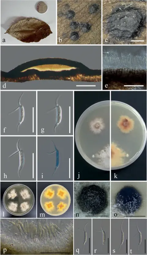
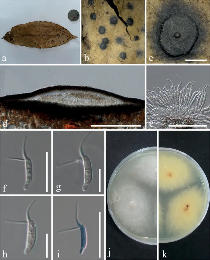
Saprobic on leaf litter. Asexual morph: Coelomycetous. Conidiomata 530–950 μm diameter (x¯ = 758 μm, n = 23), unilocular, semi-immersed, circular areas, black, mostly aggregated, sometimes solitary, with a papilla central circular ostiole (Fig. 5a–c). Conidiomata wall comprises a few to several layers of cells of textura angularis, with the inner layer being mostly thin, brown, whereas the outer layer appears dark brown to black. The longitudinal section is lenticular, the base is well developed (Fig. 5d). Conidiophores are reduced to conidiogenous cells. Conidiogenous cells enteroblastic phialidic, formed from the innermost layer of the wall, hyaline to pale brown, smooth (Fig. 5e). Conidia 11–16 × 2–3 μm (x¯ = 14 × 2.6 μm, n = 22) excluding apical appendages, mean conidium length/width ratio = 5.4:1, navicular to subcylindrical, slightly curved, 1-septate, narrow basal cell, wide middle two cells with apical cell transformed into two forked filiform cellular appendages 7–13 μm (x¯ = 10 μm, n = 22), 2–4-guttulates on the surface of the conidia, without basal appendages (Fig. 5f–i). Sexual morph: Unknown. Conidia germinated and hyphae grew in emission form the center to the outside (Fig. 5j, k). Colonies growing on PDA, reaching a diameter of 4.4 cm (n = 3) after 7 days at 26 °C, producing dense mycelium, circular, margin rough. Surface white from the surrounding of the mycelium on PDA and pale yellow in reverse.
Habitat dan peran ekologis
Saprobic on leaf litter. Asexual morph: Coelomycetous. Conidiomata 530–950 μm diameter (x¯ = 758 μm, n = 23), unilocular, semi-immersed, circular areas, black, mostly aggregated, sometimes solitary, with a papilla central circular ostiole (Fig. 5a–c). Conidiomata wall comprises a few to several layers of cells of textura angularis, with the inner layer being mostly thin, brown, whereas the outer layer appears dark brown to black. The longitudinal section is lenticular, the base is well developed (Fig. 5d). Conidiophores are reduced to conidiogenous cells. Conidiogenous cells enteroblastic phialidic, formed from the innermost layer of the wall, hyaline to pale brown, smooth (Fig. 5e). Conidia 11–16 × 2–3 μm (x¯ = 14 × 2.6 μm, n = 22) excluding apical appendages, mean conidium length/width ratio = 5.4:1, navicular to subcylindrical, slightly curved, 1-septate, narrow basal cell, wide middle two cells with apical cell transformed into two forked filiform cellular appendages 7–13 μm (x¯ = 10 μm, n = 22), 2–4-guttulates on the surface of the conidia, without basal appendages (Fig. 5f–i). Sexual morph: Unknown. Conidia germinated and hyphae grew in emission form the center to the outside (Fig. 5j, k).
- Habitat atau konteks temuan
- Informasi belum tersedia pada sumber rilis ini.
- Substrat
- Informasi belum tersedia pada sumber rilis ini.
- Peran atau konteks
- Pengurai
Reproduksi
Saprobic on leaf litter. Asexual morph: Coelomycetous. Conidiomata 530–950 μm diameter (x¯ = 758 μm, n = 23), unilocular, semi-immersed, circular areas, black, mostly aggregated, sometimes solitary, with a papilla central circular ostiole (Fig. 5a–c). Conidiomata wall comprises a few to several layers of cells of textura angularis, with the inner layer being mostly thin, brown, whereas the outer layer appears dark brown to black. The longitudinal section is lenticular, the base is well developed (Fig. 5d). Conidiophores are reduced to conidiogenous cells. Conidiogenous cells enteroblastic phialidic, formed from the innermost layer of the wall, hyaline to pale brown, smooth (Fig. 5e). Conidia 11–16 × 2–3 μm (x¯ = 14 × 2.6 μm, n = 22) excluding apical appendages, mean conidium length/width ratio = 5.4:1, navicular to subcylindrical, slightly curved, 1-septate, narrow basal cell, wide middle two cells with apical cell transformed into two forked filiform cellular appendages 7–13 μm (x¯ = 10 μm, n = 22), 2–4-guttulates on the surface of the conidia, without basal appendages (Fig. 5f–i).
Catatan identitas dan cakupan
- Nama diterima mengikuti Catalogue of Life Base2026-09-11; uraian mempertahankan konsep dan nama sumber aslinya.
- Preserve original treatment localities, hosts and substrate scope. Materials examined describe documented specimens, not the full global range.
- Impor dan terjemahan otomatis belum diperiksa editorial per profil. Struktur reproduktif yang disebutkan tidak membuktikan seluruh siklus hidup diketahui.
- Pengukuran pada uraian mengikuti spesimen, kultur, dan metode sumber; angka tidak diperluas ke semua populasi.
Catatan distribusi dalam narasi mengikuti sumbernya. Dokumentasi spesimen atau foto mengikuti catatan sumber. Bukti wilayah Indonesia dan penilaian konservasi tidak disimpulkan dari gambar.
Melihat dengan konteks
Ciliochorella savannica (IFRD:9467, holotype; IFRDCC:3201, ex-type) a, b the specimen c surface of fruiting bodies d longitudinal section of the conidioma e peridium f–h mature conidia i mature conidia in cotton blue j, k colonies on PDA (k from below). Scale bars: 400 µm (c); 200 µm (d); 20µm (e); 10 µm (f–i).
- Jenis
- Pelat ilmiah
- Kredit
- Jia-Yu Song, Hai-Xia Wu, Jin-Chen Li, Wei-Feng Ding, Cui-Ling Gong, Xiang-Yu Zeng, Nalin N. Wijayawardene, Da-Xin Yang
- Lisensi
- CC BY 4.0
- Sumber media
- Buka entri sumber
Sumber profil
- [1] Taxonomy and evolution history of two new litter-decomposing Ciliochorella (Amphisphaeriales, Sporocadaceae) — Ciliochorella savannica
- [2] Catalogue of Life Base2026-09-11 — Ciliochorella savannica
- [3] Dokumentasi visual — Ciliochorella savannica
Rujukan halaman ini
Fungi. Ciliochorella savannica. Rilis data fungi-2026-10-12-expansion-2. https://fungi-atlas.mgadib.chatgpt.site/jamur/ciliochorella-savannica/.
Status: impor sumber; pemeriksaan editorial per profil belum selesai. Untuk klaim biologis, gunakan rujukan pada bagian terkait dan periksa konteks sumber aslinya.