Ciri dan struktur
Colonies on PDA attaining 9 cm diam. in six weeks at room temperature, slow growing, dark olivaceous. Mycelium superficial and immersed composed of septate, branched, 2.3–4.5 µm wide, sub-hyaline, with smooth and thick-walled hyphae. Sexual morph Undetermined. Asexual morph Conidiophores 6–10 µm high, 3–4 µm diam. (x̄ = 8.24 × 3.52 µm, n = 10), terminal and intercalary, cylindrical or sub-cylindrical, darkened conidiogenous loci. Conidia 3–6 × 2–4 µm (x̄ = 3.64 × 2.75 µm, n = 30), forming long branched chains, hyaline to pale-olivaceous, smooth and thin-walled, aseptate, globose to ovoid with rounded ends. Colonies on PDA (Figure 2, PE58), superficial, dark olivaceous with dark-grey centre, irregular, undulate with wrinkled and raised on surface media; reverse dark olivaceous. Generative hyphae simple-septate, branched, sub-hyaline, guttules, thick-walled (Figure 8).
Habitat dan peran ekologis
Colonies on PDA attaining 9 cm diam. in six weeks at room temperature, slow growing, dark olivaceous. Mycelium superficial and immersed composed of septate, branched, 2.3–4.5 µm wide, sub-hyaline, with smooth and thick-walled hyphae. Sexual morph Undetermined. Asexual morph Conidiophores 6–10 µm high, 3–4 µm diam. (x̄ = 8.24 × 3.52 µm, n = 10), terminal and intercalary, cylindrical or sub-cylindrical, darkened conidiogenous loci. Conidia 3–6 × 2–4 µm (x̄ = 3.64 × 2.75 µm, n = 30), forming long branched chains, hyaline to pale-olivaceous, smooth and thin-walled, aseptate, globose to ovoid with rounded ends. Colonies on PDA (Figure 2, PE58), superficial, dark olivaceous with dark-grey centre, irregular, undulate with wrinkled and raised on surface media; reverse dark olivaceous. Generative hyphae simple-septate, branched, sub-hyaline, guttules, thick-walled (Figure 8).
- Habitat atau konteks temuan
- kultur yang didokumentasikan
- Substrat
- Informasi belum tersedia pada sumber rilis ini.
- Peran atau konteks
- Belum dikategorikan pada rilis ini.
Reproduksi
Colonies on PDA attaining 9 cm diam. in six weeks at room temperature, slow growing, dark olivaceous. Mycelium superficial and immersed composed of septate, branched, 2.3–4.5 µm wide, sub-hyaline, with smooth and thick-walled hyphae. Sexual morph Undetermined. Asexual morph Conidiophores 6–10 µm high, 3–4 µm diam. (x̄ = 8.24 × 3.52 µm, n = 10), terminal and intercalary, cylindrical or sub-cylindrical, darkened conidiogenous loci. Conidia 3–6 × 2–4 µm (x̄ = 3.64 × 2.75 µm, n = 30), forming long branched chains, hyaline to pale-olivaceous, smooth and thin-walled, aseptate, globose to ovoid with rounded ends.
Catatan identitas dan cakupan
- Nama diterima mengikuti Catalogue of Life Base2026-09-11; uraian mempertahankan konsep dan nama sumber aslinya.
- Preserve original treatment localities, hosts and substrate scope. Materials examined describe documented specimens, not the full global range.
- Impor dan terjemahan otomatis belum diperiksa editorial per profil. Struktur reproduktif yang disebutkan tidak membuktikan seluruh siklus hidup diketahui.
- Pengukuran pada uraian mengikuti spesimen, kultur, dan metode sumber; angka tidak diperluas ke semua populasi.
Catatan distribusi dalam narasi mengikuti sumbernya. Dokumentasi spesimen atau foto mengikuti catatan sumber. Bukti wilayah Indonesia dan penilaian konservasi tidak disimpulkan dari gambar.
Melihat dengan konteks
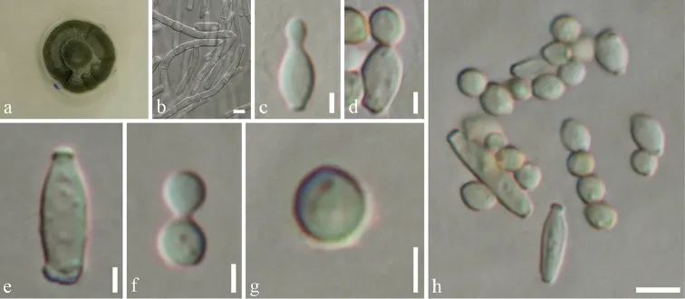
Cladosporium endophyticum (MFLU 18-0005, holotype). a Colony on MEA media b Mycelium masses c–e Conidia and conidiogenous cells f, g Conidia h Conidia and conidiogenous cells. Scale bars: 5 µm (b–h), 10 µm (h).
- Jenis
- Pelat ilmiah
- Kredit
- Saowaluck Tibpromma, Kevin D. Hyde, Jayarama D. Bhat, Peter E. Mortimer, Jianchu Xu, Itthayakorn Promputtha, Mingkwan Doilom, Jun-Bo Yang, Alvin M. C. Tang, Samantha C. Karunarathna
- Lisensi
- CC BY 4.0
- Sumber media
- Buka entri sumber
Sumber profil
- [1] Identification of endophytic fungi from leaves of Pandanaceae based on their morphotypes and DNA sequence data from southern Thailand — Cladosporium endophyticum
- [2] Catalogue of Life Base2026-09-11 — Cladosporium endophyticum
- [3] Dokumentasi visual — Cladosporium endophyticum
Rujukan halaman ini
Fungi. Cladosporium endophyticum. Rilis data fungi-2026-10-12-expansion-2. https://fungi-atlas.mgadib.chatgpt.site/jamur/cladosporium-endophyticum/.
Status: impor sumber; pemeriksaan editorial per profil belum selesai. Untuk klaim biologis, gunakan rujukan pada bagian terkait dan periksa konteks sumber aslinya.