Ciri dan struktur
Parasitic on aphids from Telosma cordata (Burm. f.) Merr. (Apocynaceae). Sexual morph: Not observed. Asexual morph: Hyphomycetous. Mycelium superficial and immersed, with abundant sporulation on the surface of aphids. Conidiophores erect, fasciculate, macronematous, cylindrical, subnodulose or nodulose, geniculate, septate, branched, pale brown, slightly roughened to verruculose, thick-walled, and pronounced loci, 111–367 × 3.5–5 μm. Conidiogenous cells integrated, terminal or intercalary, cylindrical, cylindrical-oblong, sometimes geniculate, 20–65 × 3–6 μm, conidiogenous loci at the apex (2–5) or in lateral shoulders (0–4). Ramoconidia olive-green, 0–4-septate, ellipsoidal to subcylindrical, smooth- and thick-walled, 10–26 × 3.5–7.5 μm. Secondary ramoconidia pale brown, oblong, oblong-ellipsoid, 0–1septate, 1–4 distal hila, 8.5–13.5 × 3.5–7.5 μm. Conidia numerous, catenate, forming short branched chains, aseptate, olive to light olive, smooth- and thin-walled, variable in size and shape, ellipsoid-ovoid, obovoid, and fusiform, 4–10 × 3–6.5 μm. Intercalary conidia aseptate, olive to light olive, ellipsoid-ovoid, fusiform, 6–10 × 4–6.5 μm. Terminal conidia aseptate, olive to light olive, obovoid, 4–6.5 × 3–5.5 μm. Microcyclic conidiogenesis absent. In vitro on SNA: Mycelium abundant, submerged, overgrowing whole culture dishes, hyphae straight to slightly sinuous, septate and branched, light olive-green to olive-green, and thick-walled, 1.5–4.5 µm wide. Conidiophores erect, branched, light olive-green, thick-walled, 43–163 × 2.5–4 µm. Ramoconidia pale olivegreen, narrowly ellipsoid to cylindrical-oblong, subcylindrical, septate or aseptate, smooth- and thick-walled, 6–27 × 3–5 µm.
Habitat dan peran ekologis
Aphids (Aphididae), leaves of plants, and uredinia of Hemileia vastatrix (Pucciniaceae). In tropical and subtropical regions, it can infect insects, act saprophytically on plant tissues, and function as a fungicolous fungus on the uredinia of rust. During the middle and late stages of aphid damage, numerous aphids die on the underside of plant leaves, becoming enveloped in mycelial tissue. China, Sichuan Province, Guangan City. China, Guizhou Province, Guiyang City. Ethiopia, Oromia Region, Illubabor Gore. Brazil, Minas Gerais, Viçosa.
- Habitat atau konteks temuan
- Informasi belum tersedia pada sumber rilis ini.
- Substrat
- Informasi belum tersedia pada sumber rilis ini.
- Peran atau konteks
- Belum dikategorikan pada rilis ini.
Reproduksi
Parasitic on aphids from Telosma cordata (Burm. f.) Merr. (Apocynaceae). Sexual morph: Not observed. Asexual morph: Hyphomycetous. Mycelium superficial and immersed, with abundant sporulation on the surface of aphids. Conidiophores erect, fasciculate, macronematous, cylindrical, subnodulose or nodulose, geniculate, septate, branched, pale brown, slightly roughened to verruculose, thick-walled, and pronounced loci, 111–367 × 3.5–5 μm. Conidiogenous cells integrated, terminal or intercalary, cylindrical, cylindrical-oblong, sometimes geniculate, 20–65 × 3–6 μm, conidiogenous loci at the apex (2–5) or in lateral shoulders (0–4). Ramoconidia olive-green, 0–4-septate, ellipsoidal to subcylindrical, smooth- and thick-walled, 10–26 × 3.5–7.5 μm.
Catatan identitas dan cakupan
- Nama diterima mengikuti Catalogue of Life Base2026-09-11; uraian mempertahankan konsep dan nama sumber aslinya.
- Preserve original treatment localities, hosts and substrate scope. Materials examined describe documented specimens, not the full global range.
- Impor dan terjemahan otomatis belum diperiksa editorial per profil. Struktur reproduktif yang disebutkan tidak membuktikan seluruh siklus hidup diketahui.
- Pengukuran pada uraian mengikuti spesimen, kultur, dan metode sumber; angka tidak diperluas ke semua populasi.
Catatan distribusi dalam narasi mengikuti sumbernya. Dokumentasi spesimen atau foto mengikuti catatan sumber. Bukti wilayah Indonesia dan penilaian konservasi tidak disimpulkan dari gambar.
Melihat dengan konteks
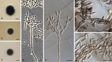
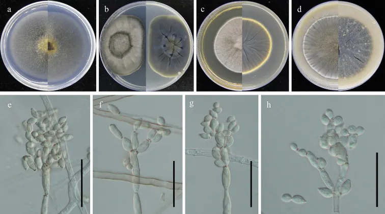
Cladosporium guizhouense (GUCC 401.7). a–d colony on SNA, PDA, MEA and OA (left: above, right: reverse) e–h conidiogenous cells, secondary ramoconidia and conidia on SNA. Scale bars: 10 µm (e–h).
- Jenis
- Pelat ilmiah
- Kredit
- Si-Yao Wang, Yong Wang, Yan Li
- Lisensi
- CC BY 4.0
- Sumber media
- Buka entri sumber
Dokumentasi tambahan
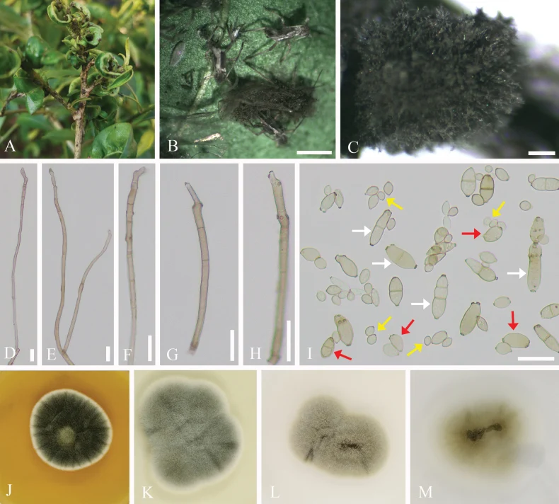
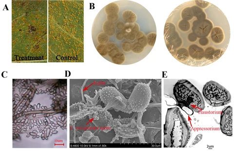
Cladosporium guizhouense. A. Symptoms observed on host. B, C. Appearance of colonies on aphids. D–H. Conidiophores. I. Ramoconidia (white arrows), secondary ramoconidia (red arrows), and terminal conidia (yellow arrows). J–M. Colonies on MEA, PDA, OA, and SNA for 7 days. Scale bars: 1000 µm (B); 200 µm (C); 20 µm (D–I).
Kredit dan penggunaan
Chunlin Yang, Xiulan Xu, Xinyue Li, Feng Liu, Zhen Zeng, Qiangang Xiao, Yinggao Liu
Ukuran dan format gambar disesuaikan tanpa pemotongan, retus, atau penambahan skala. Lisensi sumber dipertahankan.
Sumber profil
- [1] Morphological and phylogenetic analyses reveal novel entomopathogenic fungi infecting scale insects and aphids in China — Cladosporium guizhouense
- [2] Catalogue of Life Base2026-09-11 — Cladosporium guizhouense
- [3] Dokumentasi visual — Cladosporium guizhouense
- [4] Dokumentasi visual — Cladosporium guizhouense
Rujukan halaman ini
Fungi. Cladosporium guizhouense. Rilis data fungi-2026-10-12-expansion-2. https://fungi-atlas.mgadib.chatgpt.site/jamur/cladosporium-guizhouense/.
Status: impor sumber; pemeriksaan editorial per profil belum selesai. Untuk klaim biologis, gunakan rujukan pada bagian terkait dan periksa konteks sumber aslinya.