Ciri dan struktur
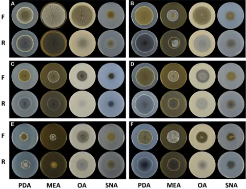
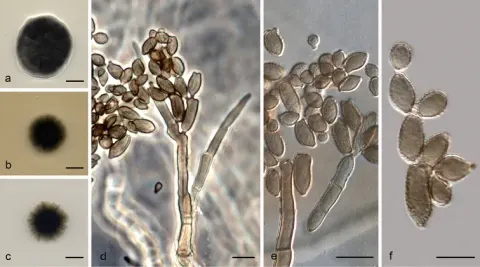
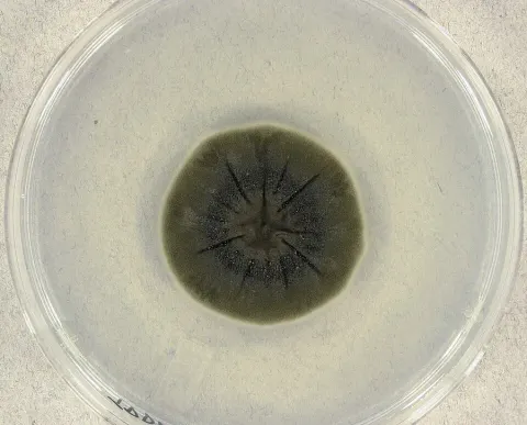
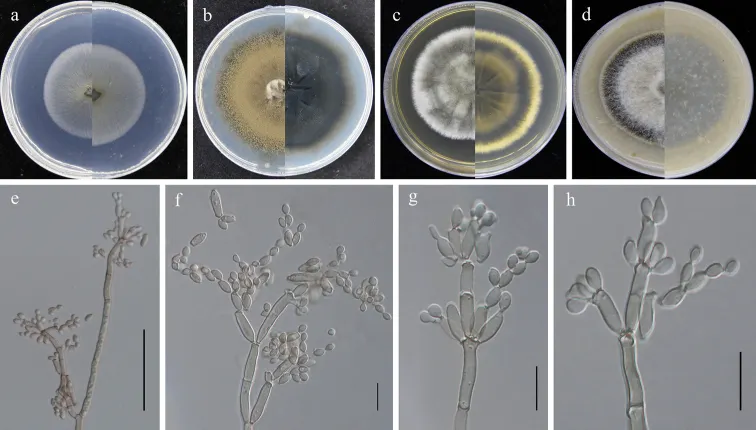
Saprobic on fallen leaves of Eucommia ulmoides. Sexual morph: Not developed. Asexual morph: Hyphomycetous. Mycelium superficial, hyphae branched, hyaline to subhyaline, 2.5–5 μm wide, usually slightly constricted at the septa and somewhat swollen, smooth to somewhat verruculose or irregularly rough-walled. Conidiophores macro- and micronematous, 14–167 × 2.5–4.5 µm (x̄ = 65.4 × 3.4 µm; n = 20), erect, branched, slightly attenuated towards the apex, light olive-green, smooth and thick-walled. Conidia in acropetal chains, 2–9.5 × 2–4 µm (x̄ = 5.6 × 3.3 µm; n = 30) mostly light olive-green, aseptate, mostly smooth-walled and thin-walled, variable in size and shape, subglobose, ellipsoid-ovoid, obovoid, fusiform. Secondary ramoconidia 6–24 × 2–5.5 µm (x̄ = 11.2 × 3.3 µm; n = 30), olive-green, narrowly ellipsoid to cylindrical-oblong, subcylindrical, aseptate, rarely 1-septate, mostly smooth-walled and thick-walled. Colonies on SNA 30–40 mm diam, after 2 weeks at 25 °C, pale olive to pale whitish, flat, velvety, with a regular edge, reverse light olive to light white. Colonies on PDA 30–40 mm diam, after 2 weeks at 25 °C, olive-gray to olive-green or olive-brown, powdery or flocculent, fluffy, regular, radially furrowed, lacerated or feathery, and often forming a gray-white or olive bulge in the colony kernel; reverse dark olive or dull green to black. Colonies on MEA 35–45 mm diam, after 2 weeks at 25 °C, gray-green to white or gray-white, fluffy, radially furrowed, with a whitish final edge; reverse olive-yellow to olive-gray to olive-green, with a whitish final edge. Colonies on OA 35–45 mm diam, after 2 weeks at 25°C, white to olive-green, with a pale gray final edge, velvety or fluffy, margins colorless or pale gray, glabrous, regular; reverse olive-green to dark green.
Habitat dan peran ekologis
Saprobic on fallen leaves of Eucommia ulmoides. Sexual morph: Not developed. Asexual morph: Hyphomycetous. Mycelium superficial, hyphae branched, hyaline to subhyaline, 2.5–5 μm wide, usually slightly constricted at the septa and somewhat swollen, smooth to somewhat verruculose or irregularly rough-walled. Conidiophores macro- and micronematous, 14–167 × 2.5–4.5 µm (x̄ = 65.4 × 3.4 µm; n = 20), erect, branched, slightly attenuated towards the apex, light olive-green, smooth and thick-walled. Conidia in acropetal chains, 2–9.5 × 2–4 µm (x̄ = 5.6 × 3.3 µm; n = 30) mostly light olive-green, aseptate, mostly smooth-walled and thin-walled, variable in size and shape, subglobose, ellipsoid-ovoid, obovoid, fusiform. Secondary ramoconidia 6–24 × 2–5.5 µm (x̄ = 11.2 × 3.3 µm; n = 30), olive-green, narrowly ellipsoid to cylindrical-oblong, subcylindrical, aseptate, rarely 1-septate, mostly smooth-walled and thick-walled. Colonies on SNA 30–40 mm diam, after 2 weeks at 25 °C, pale olive to pale whitish, flat, velvety, with a regular edge, reverse light olive to light white. Colonies on PDA 30–40 mm diam, after 2 weeks at 25 °C, olive-gray to olive-green or olive-brown, powdery or flocculent, fluffy, regular, radially furrowed, lacerated or feathery, and often forming a gray-white or olive bulge in the colony kernel; reverse dark olive or dull green to black.
- Habitat atau konteks temuan
- kultur yang didokumentasikan
- Substrat
- Informasi belum tersedia pada sumber rilis ini.
- Peran atau konteks
- Pengurai
Reproduksi
Saprobic on fallen leaves of Eucommia ulmoides. Sexual morph: Not developed. Asexual morph: Hyphomycetous. Mycelium superficial, hyphae branched, hyaline to subhyaline, 2.5–5 μm wide, usually slightly constricted at the septa and somewhat swollen, smooth to somewhat verruculose or irregularly rough-walled. Conidiophores macro- and micronematous, 14–167 × 2.5–4.5 µm (x̄ = 65.4 × 3.4 µm; n = 20), erect, branched, slightly attenuated towards the apex, light olive-green, smooth and thick-walled. Conidia in acropetal chains, 2–9.5 × 2–4 µm (x̄ = 5.6 × 3.3 µm; n = 30) mostly light olive-green, aseptate, mostly smooth-walled and thin-walled, variable in size and shape, subglobose, ellipsoid-ovoid, obovoid, fusiform.
Catatan identitas dan cakupan
- Nama diterima mengikuti Catalogue of Life Base2026-09-11; uraian mempertahankan konsep dan nama sumber aslinya.
- Preserve original treatment localities, hosts and substrate scope. Materials examined describe documented specimens, not the full global range.
- Impor dan terjemahan otomatis belum diperiksa editorial per profil. Struktur reproduktif yang disebutkan tidak membuktikan seluruh siklus hidup diketahui.
- Pengukuran pada uraian mengikuti spesimen, kultur, dan metode sumber; angka tidak diperluas ke semua populasi.
Catatan distribusi dalam narasi mengikuti sumbernya. Dokumentasi spesimen atau foto mengikuti catatan sumber. Bukti wilayah Indonesia dan penilaian konservasi tidak disimpulkan dari gambar.
Melihat dengan konteks
Cladosporium perangustum (GUCC 401.6, new substrate record from Guizhou Province). a–d colonies on SNA, PDA, MEA and OA (left: above, right: reverse) e branching conidiophore, secondary ramoconidia and conidia on SNA f–h conidiogenous cells, secondary ramoconidia and conidia on SNA. Scale bars: 50 µm (e); 10 µm (f–h).
- Jenis
- Pelat ilmiah
- Kredit
- Si-Yao Wang, Yong Wang, Yan Li
- Lisensi
- CC BY 4.0
- Sumber media
- Buka entri sumber
Sumber profil
- [1] Cladosporium spp. (Cladosporiaceae) isolated from Eucommia ulmoides in China — Cladosporium perangustum
- [2] Catalogue of Life Base2026-09-11 — Cladosporium perangustum
- [3] Dokumentasi visual — Cladosporium perangustum
Rujukan halaman ini
Fungi. Cladosporium perangustum. Rilis data fungi-2026-10-12-expansion-2. https://fungi-atlas.mgadib.chatgpt.site/jamur/cladosporium-perangustum/.
Status: impor sumber; pemeriksaan editorial per profil belum selesai. Untuk klaim biologis, gunakan rujukan pada bagian terkait dan periksa konteks sumber aslinya.