Ciri dan struktur
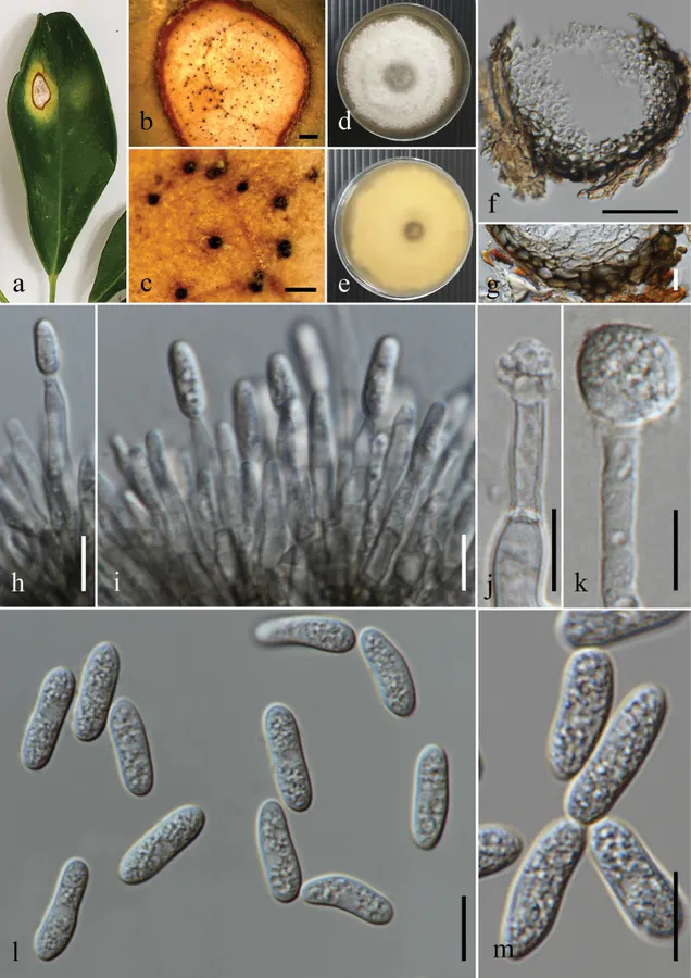
Associated with leaf spots of Terminalia sp. Leaf spots irregular or oval, pale brown, surrounded with a dark brown margin. Sexual morph: Not observed. Asexual morph on substrate: Conidiomata 70–120 × 60–120 µm (x̄ = 94.5 × 96 µm, n = 10), semi-immersed, scattered, globose to subglobose, black, sometimes erumpent. Setae not observed. Conidiomatal wall 8.5–21 µm thick (x̄ = 14.6 µm, n = 10), consisting of 3–5 layers of thick-walled pseudoparenchymatous cells of textura angularis, outer layers dark brown, inner layer pale brown to hyaline. Asexual morph on PDA: Conidiophores hyaline, smooth-walled, aseptate, unbranched. Conidiogenous cells 16–22 × 3–4.5 µm (x̄ = 18.9 × 3.6 µm, n = 10), hyaline, cylindrical to ampulliform, straight or flexuous, tapering towards the apex. Conidia 13–19 × 4.5–6 µm (x̄ = 15.8 × 5.1 µm, n = 30; L/W ratio = 3.1), hyaline, cylindrical or oblong, smooth-walled, guttulate, aseptate, mostly with rounded ends. Appressoria 7–12 × 7–13 µm (x̄ = 10.9 × 10.5 µm, n = 5), hyaline, single-celled, globose to subglobose or irregular, smooth-walled. Colonies on PDA reaching approximately 45 mm diam. after 7 d of incubation at 25 °C; mycelium greyish white, elevation flat or raised, aerial and filamentous with an entire or undulate margin.
Habitat dan peran ekologis
(listed chronologically). Endophytic on Theobroma cacao and Genipa americana in Panama (Rojas et al. 2010) and Terpsichore taxifolia in Puerto Rico (Doyle et al. 2013). Pathogenic; anthracnose on fruits of Musa sp. in Minas Gerais, Pernambuco, Santa Catarina, and São Paulo states in Brazil and Mexico (Vieira et al. 2017; Fuentes-Aragón et al. 2021), leaves of Anacardium humile and A. occidentale in Brazil (Veloso et al. 2018, 2021), fruits of Mangifera indica in Mexico (Fuentes-Aragón et al. 2020a), fruits of Persea americana in Mexico (Fuentes-Aragón et al. 2020b) and Brazil (Soares et al. 2021), leaves of Bauhinia forficata in Brazil (de Souza Junior et al. 2021), leaves of Manihot esculenta in Brazil (Machado et al. 2021), fruits of Carica papaya in Mexico (Pacheco-Esteva et al. 2022), and leaves of V accinium corymbosum in Brazil (Soares et al. 2022). Bitter rot of Malus domestica in New York (Khodadadi et al. 2020) and Spain (Cabrefiga et al. 2022). Preharvest decay of Malus domestica in Italy (Deltedesco and Oettl 2023). Leaf spots on Euterpe oleracea and Malus domestica in Brazil and Uruguay (Astolfi et al. 2022; dos Santos et al. 2022; Andrello et al. 2024). Associated with leaf spots of Terminalia sp. in Thailand (this study).
- Habitat atau konteks temuan
- Informasi belum tersedia pada sumber rilis ini.
- Substrat
- Informasi belum tersedia pada sumber rilis ini.
- Peran atau konteks
- Belum dikategorikan pada rilis ini.
Reproduksi
Associated with leaf spots of Terminalia sp. Leaf spots irregular or oval, pale brown, surrounded with a dark brown margin. Sexual morph: Not observed. Asexual morph on substrate: Conidiomata 70–120 × 60–120 µm (x̄ = 94.5 × 96 µm, n = 10), semi-immersed, scattered, globose to subglobose, black, sometimes erumpent. Setae not observed. Conidiomatal wall 8.5–21 µm thick (x̄ = 14.6 µm, n = 10), consisting of 3–5 layers of thick-walled pseudoparenchymatous cells of textura angularis, outer layers dark brown, inner layer pale brown to hyaline. Asexual morph on PDA: Conidiophores hyaline, smooth-walled, aseptate, unbranched. Conidiogenous cells 16–22 × 3–4.5 µm (x̄ = 18.9 × 3.6 µm, n = 10), hyaline, cylindrical to ampulliform, straight or flexuous, tapering towards the apex.
Catatan identitas dan cakupan
- Nama diterima mengikuti Catalogue of Life Base2026-09-11; uraian mempertahankan konsep dan nama sumber aslinya.
- Preserve original treatment localities, hosts and substrate scope. Materials examined describe documented specimens, not the full global range.
- Impor dan terjemahan otomatis belum diperiksa editorial per profil. Struktur reproduktif yang disebutkan tidak membuktikan seluruh siklus hidup diketahui.
- Pengukuran pada uraian mengikuti spesimen, kultur, dan metode sumber; angka tidak diperluas ke semua populasi.
Catatan distribusi dalam narasi mengikuti sumbernya. Dokumentasi spesimen atau foto mengikuti catatan sumber. Bukti wilayah Indonesia dan penilaian konservasi tidak disimpulkan dari gambar.
Melihat dengan konteks
Colletotrichum chrysophilum. a. Leaf spot (Terminalia sp.); b. Close up of a leaf spot; c. Conidiomata on substrate; d, e. Top and reverse of colony on PDA after 3 d f Section through a conidioma; g. Conidiomatal wall; h, i. Conidiogenesis and developing conidia; j, k. Appressoria; l, m. Conidia. Scale bars: 1 mm (b); 200 μm (c); 50 μm (f); 10 μm (g–m).
- Jenis
- Pelat ilmiah
- Kredit
- Deecksha Gomdola, Rajesh Jeewon, Eric H.C. McKenzie, Ruvishika S. Jayawardena, Fatimah Al-Otibi, Xia Tang, Yong Wang, Kevin D. Hyde, Li Fu
- Lisensi
- CC BY 4.0
- Sumber media
- Buka entri sumber
Sumber profil
- [1] Phylogenetic diversity of Colletotrichum species (Sordariomycetes, Glomerellales, Glomerellaceae) associated with plant diseases in Thailand — Colletotrichum chrysophilum
- [2] Catalogue of Life Base2026-09-11 — Colletotrichum chrysophilum
- [3] Dokumentasi visual — Colletotrichum chrysophilum
Rujukan halaman ini
Fungi. Colletotrichum chrysophilum. Rilis data fungi-2026-10-12-expansion-2. https://fungi-atlas.mgadib.chatgpt.site/jamur/colletotrichum-chrysophilum/.
Status: impor sumber; pemeriksaan editorial per profil belum selesai. Untuk klaim biologis, gunakan rujukan pada bagian terkait dan periksa konteks sumber aslinya.