01 / Morfologi

Ciri dan struktur

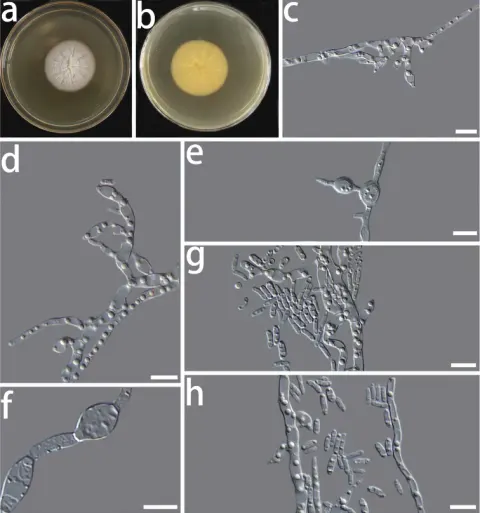
Colony on PDA after 8 d, hyphae hyaline, multi-guttulate, septate, smooth-walled; conidiophores short; conidiogenous cells hyaline, phialidic or oval in shape, single or in clusters on short lateral branches, measuring 2.9–7.2 × 1.8–3.7 μm (x̄= 4.7 × 2.6 μm, n = 50) (Fig. 2f, g); two types of conidia were observed, swollen conidia were hyaline, one-celled, dumb-bell-shaped, with hyphae emerging from both ends (Fig. 2d, e), measuring 7.6–16.5 × 2.3–4.1 μm (x̄ = 9.7 × 3.1 μm, n = 50) (Fig. 2c), oblong conidia were hyaline, one-celled, often oblong to ellipsoidal in shape, measuring 3.4–6.8 × 1.4–2.7 μm (x̄ = 4.7 × 1.8 μm, n = 50) (Fig. 2h). Chlamydospores absent. Sexual morph unknown. The optimal temperature for growth was 25 °C on PDA. No growth was detected at 5 and 35 °C. Colonies on PDA after 8 d at 25 °C were white, circular, margin entire, flat, dense, partially immersed in the medium and sticky protuberance at the centre of the colony.

Sebagian uraian ditampilkan dalam bahasa sumber; terjemahannya belum tersedia atau belum lolos pemeriksaan.

Rujukan: [1]

Pengukuran mengikuti objek, spesimen atau kultur, dan metode yang disebutkan sumber. Baca uraian asli untuk konteks lengkap.

02 / Kehidupan

Habitat dan peran ekologis

China, Yunnan Province: Tiesuo township, 26°32'71"N, 100°57'3"E, ca. 2120 m elev., isolated from Candelaria fibrosa, 13 Nov 2020, H. L. Si, CX03C1 (HMAS 350271, holotype), ex-type culture CGMCC3.20304. Colony on PDA after 8 d, hyphae hyaline, multi-guttulate, septate, smooth-walled; conidiophores short; conidiogenous cells hyaline, phialidic or oval in shape, single or in clusters on short lateral branches, measuring 2.9–7.2 × 1.8–3.7 μm (x̄= 4.7 × 2.6 μm, n = 50) (Fig. 2f, g); two types of conidia were observed, swollen conidia were hyaline, one-celled, dumb-bell-shaped, with hyphae emerging from both ends (Fig. 2d, e), measuring 7.6–16.5 × 2.3–4.1 μm (x̄ = 9.7 × 3.1 μm, n = 50) (Fig. 2c), oblong conidia were hyaline, one-celled, often oblong to ellipsoidal in shape, measuring 3.4–6.8 × 1.4–2.7 μm (x̄ = 4.7 × 1.8 μm, n = 50) (Fig. 2h). Chlamydospores absent. Sexual morph unknown. The optimal temperature for growth was 25 °C on PDA. No growth was detected at 5 and 35 °C. Colonies on PDA after 8 d at 25 °C were white, circular, margin entire, flat, dense, partially immersed in the medium and sticky protuberance at the centre of the colony. China, Yunnan Province: Tiesuo township, 26°32'71"N, 100°57'3"E, ca. 2120 m elev., isolated from on Candelaria fibrosa, 13 Nov 2020, H.

Sebagian uraian ditampilkan dalam bahasa sumber; terjemahannya belum tersedia atau belum lolos pemeriksaan.

Rujukan: [1]

Habitat atau konteks temuan
kultur yang didokumentasikan
Substrat
Informasi belum tersedia pada sumber rilis ini.
Peran atau konteks
Belum dikategorikan pada rilis ini.
Temukan hubungan serupa
03 / Siklus hidup

Reproduksi

Colony on PDA after 8 d, hyphae hyaline, multi-guttulate, septate, smooth-walled; conidiophores short; conidiogenous cells hyaline, phialidic or oval in shape, single or in clusters on short lateral branches, measuring 2.9–7.2 × 1.8–3.7 μm (x̄= 4.7 × 2.6 μm, n = 50) (Fig. 2f, g); two types of conidia were observed, swollen conidia were hyaline, one-celled, dumb-bell-shaped, with hyphae emerging from both ends (Fig. 2d, e), measuring 7.6–16.5 × 2.3–4.1 μm (x̄ = 9.7 × 3.1 μm, n = 50) (Fig. 2c), oblong conidia were hyaline, one-celled, often oblong to ellipsoidal in shape, measuring 3.4–6.8 × 1.4–2.7 μm (x̄ = 4.7 × 1.8 μm, n = 50) (Fig. 2h). Chlamydospores absent. Sexual morph unknown.

Sebagian uraian ditampilkan dalam bahasa sumber; terjemahannya belum tersedia atau belum lolos pemeriksaan.

Rujukan: [1]

04 / Batas informasi

Catatan identitas dan cakupan

  • Nama diterima mengikuti Catalogue of Life Base2026-09-11; uraian mempertahankan konsep dan nama sumber aslinya.
  • Preserve original treatment localities, hosts and substrate scope. Materials examined describe documented specimens, not the full global range.
  • Impor dan terjemahan otomatis belum diperiksa editorial per profil. Struktur reproduktif yang disebutkan tidak membuktikan seluruh siklus hidup diketahui.
  • Pengukuran pada uraian mengikuti spesimen, kultur, dan metode sumber; angka tidak diperluas ke semua populasi.

Catatan distribusi dalam narasi mengikuti sumbernya. Dokumentasi spesimen atau foto mengikuti catatan sumber. Bukti wilayah Indonesia dan penilaian konservasi tidak disimpulkan dari gambar.

05 / Dokumentasi

Melihat dengan konteks

Morphological characters of Coniochaeta fibrosae sp. nov. (HMAS 350271) a, b cultures on PDA from the surface and reverse c swollen conidia d, e swollen conidia germinate hyphae f, g conidiogenous cells h conidia. Scale bars: 10 μm.

Jenis
Pelat ilmiah
Kredit
Hong-Li Si, Yue-Min Su, Xiao-Xiao Zheng, Meng-Yao Ding, Tanay Bose, Run-Lei Chang
Lisensi
CC BY 4.0
Sumber media
Buka entri sumber

Gambar menunjukkan objek menurut identifikasi sumber, bukan verifikasi independen spesimen oleh Fungi. Tidak ada skala baru yang ditambahkan.

06 / Rujukan

Sumber profil

  1. [1] Phylogenetic and morphological analyses of Coniochaeta isolates recovered from Inner Mongolia and Yunnan revealed three new endolichenic fungal species — Coniochaeta fibrosaeBagian sumber: PMC8445908_section_11
    Tanggal penghimpunan sumber: 2026-10-12
    Teks sumber: CC BY 4.0 · Si HL, Su YM, Zheng XX, Ding MY, Bose T, Chang RL.
  2. [2] Catalogue of Life Base2026-09-11 — Coniochaeta fibrosaeBagian sumber: ColDP NameUsage.tsv ID 9Z4Q8; accepted species; source 2073
    Tanggal penghimpunan sumber: 2026-10-12
  3. [3] Dokumentasi visual — Coniochaeta fibrosaeBagian sumber: Individual media record; identification follows the source, not independent determination
    Tanggal penghimpunan sumber: 2026-10-12
    Media sumber: CC BY 4.0 · Hong-Li Si, Yue-Min Su, Xiao-Xiao Zheng, Meng-Yao Ding, Tanay Bose, Run-Lei Chang

Adaptasi teks: CC BY 4.0. Si HL, Su YM, Zheng XX, Ding MY, Bose T, Chang RL. — Phylogenetic and morphological analyses of Coniochaeta isolates recovered from Inner Mongolia and Yunnan revealed three new endolichenic fungal species — Coniochaeta fibrosae

Uraian asli dan riwayat sumber

Riwayat atau halaman sumber

Ringkasan sumber

Colony on PDA after 8 d, hyphae hyaline, multi-guttulate, septate, smooth-walled; conidiophores short; conidiogenous cells hyaline, phialidic or oval in shape, single or in clusters on short lateral branches, measuring 2.9–7.2 × 1.8–3.7 μm (x̄= 4.7 × 2.6 μm, n = 50) (Fig. 2f, g); two types of conidia were observed, swollen conidia were hyaline, one-celled, dumb-bell-shaped, with hyphae emerging from both ends (Fig. 2d, e), measuring 7.6–16.5 × 2.3–4.1 μm (x̄ = 9.7 × 3.1 μm, n = 50) (Fig. 2c), oblong conidia were hyaline, one-celled, often oblong to ellipsoidal in shape, measuring 3.4–6.8 × 1.4–2.7 μm (x̄ = 4.7 × 1.8 μm, n = 50) (Fig. 2h).

Revisi sumber

Morfologi sumber

Colony on PDA after 8 d, hyphae hyaline, multi-guttulate, septate, smooth-walled; conidiophores short; conidiogenous cells hyaline, phialidic or oval in shape, single or in clusters on short lateral branches, measuring 2.9–7.2 × 1.8–3.7 μm (x̄= 4.7 × 2.6 μm, n = 50) (Fig. 2f, g); two types of conidia were observed, swollen conidia were hyaline, one-celled, dumb-bell-shaped, with hyphae emerging from both ends (Fig. 2d, e), measuring 7.6–16.5 × 2.3–4.1 μm (x̄ = 9.7 × 3.1 μm, n = 50) (Fig. 2c), oblong conidia were hyaline, one-celled, often oblong to ellipsoidal in shape, measuring 3.4–6.8 × 1.4–2.7 μm (x̄ = 4.7 × 1.8 μm, n = 50) (Fig. 2h). Chlamydospores absent. Sexual morph unknown. The optimal temperature for growth was 25 °C on PDA. No growth was detected at 5 and 35 °C. Colonies on PDA after 8 d at 25 °C were white, circular, margin entire, flat, dense, partially immersed in the medium and sticky protuberance at the centre of the colony.

Revisi sumber

Ekologi sumber

China, Yunnan Province: Tiesuo township, 26°32'71"N, 100°57'3"E, ca. 2120 m elev., isolated from Candelaria fibrosa, 13 Nov 2020, H. L. Si, CX03C1 (HMAS 350271, holotype), ex-type culture CGMCC3.20304. Colony on PDA after 8 d, hyphae hyaline, multi-guttulate, septate, smooth-walled; conidiophores short; conidiogenous cells hyaline, phialidic or oval in shape, single or in clusters on short lateral branches, measuring 2.9–7.2 × 1.8–3.7 μm (x̄= 4.7 × 2.6 μm, n = 50) (Fig. 2f, g); two types of conidia were observed, swollen conidia were hyaline, one-celled, dumb-bell-shaped, with hyphae emerging from both ends (Fig. 2d, e), measuring 7.6–16.5 × 2.3–4.1 μm (x̄ = 9.7 × 3.1 μm, n = 50) (Fig. 2c), oblong conidia were hyaline, one-celled, often oblong to ellipsoidal in shape, measuring 3.4–6.8 × 1.4–2.7 μm (x̄ = 4.7 × 1.8 μm, n = 50) (Fig. 2h). Chlamydospores absent. Sexual morph unknown. The optimal temperature for growth was 25 °C on PDA. No growth was detected at 5 and 35 °C. Colonies on PDA after 8 d at 25 °C were white, circular, margin entire, flat, dense, partially immersed in the medium and sticky protuberance at the centre of the colony. China, Yunnan Province: Tiesuo township, 26°32'71"N, 100°57'3"E, ca. 2120 m elev., isolated from on Candelaria fibrosa, 13 Nov 2020, H.

Revisi sumber

Struktur reproduksi sumber

Colony on PDA after 8 d, hyphae hyaline, multi-guttulate, septate, smooth-walled; conidiophores short; conidiogenous cells hyaline, phialidic or oval in shape, single or in clusters on short lateral branches, measuring 2.9–7.2 × 1.8–3.7 μm (x̄= 4.7 × 2.6 μm, n = 50) (Fig. 2f, g); two types of conidia were observed, swollen conidia were hyaline, one-celled, dumb-bell-shaped, with hyphae emerging from both ends (Fig. 2d, e), measuring 7.6–16.5 × 2.3–4.1 μm (x̄ = 9.7 × 3.1 μm, n = 50) (Fig. 2c), oblong conidia were hyaline, one-celled, often oblong to ellipsoidal in shape, measuring 3.4–6.8 × 1.4–2.7 μm (x̄ = 4.7 × 1.8 μm, n = 50) (Fig. 2h). Chlamydospores absent. Sexual morph unknown.

Revisi sumber

Rujukan halaman ini

Fungi. Coniochaeta fibrosae. Rilis data fungi-2026-10-12-expansion-2. https://fungi-atlas.mgadib.chatgpt.site/jamur/coniochaeta-fibrosae/.

Status: impor sumber; pemeriksaan editorial per profil belum selesai. Untuk klaim biologis, gunakan rujukan pada bagian terkait dan periksa konteks sumber aslinya.