Ciri dan struktur
Apothecia not confluent regularly circular to irregular from the top view, at first oblate, then turbinate, finally pulvinate from the side view, sessile, subhyaline to creamy grey or pale yellowish, glabrous, * 170–420 µm in diameter, solitary or gregarious. Hymenium only very finely scurfy, ascal protrusions not clearly visible. Margin rounded in vertical median section, entire and smooth, expanded with downwards positioned rim, never raised above hymenial plane. Outer surface smooth, concolorous with the hymenium. Subicular hyphae indistinguishable. Hymenium * 75–98 µm thick. Asci shortly cylindric with slightly truncate apex, * 60–74.8 × 13.4–15.5 µm, † 51.5–62 × 11.8–14 µm (Q = 3.8–5.2), when mature * protruding above hymenium up to 7.5 µm, pars sporifera * 28–34 µm, 8-spored, hyaline; base attenuated, bifurcate, arising from perforated crosiers; only optimally oriented fully mature asci with flat lentiform operculum clearly delimited prior the spore discharge, * 6.3–6.6 µm in diam. and * 0.5 µm thick, lateral wall 3-layered, * 0.6 µm thick, after spore discharge operculum as a rule clearly visible; in IKI inamyloid; in CR outermost wall vividly rutile-red throughout the ascal length, median layer pale rutile-yellow, innermost layer greyish; in CB asci cyanophobic. Ascospores * 7.9– 8.8 –9.6 × 4.8– 5.2 –5.6 µm, † 8– 9.1 –9.5 × 4.2– 5 –5.2 µm, * Q = 1.5– 1.6 –1.9, † Q = 1.6– 1.9 –2.0, bilaterally symmetrical with one side flattened, subphaseoliform to phaseoliform, poles rounded, 1-celled; uni- to biseriate in living asci, freshly ejected remain in a group for a while due to the delicate subglobose sticky sheath enveloping individual spores; hyaline, smooth; wall 3-layered, 0.4 µm thick, perispore dull, epispore brightly refractive, endospore subhyaline, barely optically differentiated; eguttulate, uninucleate, nucleus always ±polarly positioned, 2.2–2.5 µm wide; in IKI perispore and epispore not stained, endospore purplish, nucleus slightly contrasted; in CRB without differential stainings, the edges of spore sheath sharply contrasted, after applying KOH spore sheath instantly dissolves, perispore not loosening, endospore layer purplish-rosaceous; in CB with one eccentrically positioned de Bary bubble in mature spores, perispore not loosening, moderately cyanophilic.
Habitat dan peran ekologis
The species is known so far only from Mt. Velebit, Croatia. The only collection originates from chamois dung in the alpine karstic habitat.
- Habitat atau konteks temuan
- Informasi belum tersedia pada sumber rilis ini.
- Substrat
- Informasi belum tersedia pada sumber rilis ini.
- Peran atau konteks
- Belum dikategorikan pada rilis ini.
Reproduksi
Apothecia not confluent regularly circular to irregular from the top view, at first oblate, then turbinate, finally pulvinate from the side view, sessile, subhyaline to creamy grey or pale yellowish, glabrous, * 170–420 µm in diameter, solitary or gregarious. Hymenium only very finely scurfy, ascal protrusions not clearly visible. Margin rounded in vertical median section, entire and smooth, expanded with downwards positioned rim, never raised above hymenial plane. Outer surface smooth, concolorous with the hymenium. Subicular hyphae indistinguishable. Hymenium * 75–98 µm thick. Asci shortly cylindric with slightly truncate apex, * 60–74.8 × 13.4–15.5 µm, † 51.5–62 × 11.8–14 µm (Q = 3.8–5.2), when mature * protruding above hymenium up to 7.5 µm, pars sporifera * 28–34 µm, 8-spored, hyaline; base attenuated, bifurcate, arising from perforated crosiers; only optimally oriented fully mature asci with flat lentiform operculum clearly delimited prior the spore discharge, * 6.3–6.6 µm in diam. and * 0.5 µm thick, lateral wall 3-layered, * 0.6 µm thick, after spore discharge operculum as a rule clearly visible; in IKI inamyloid; in CR outermost wall vividly rutile-red throughout the ascal length, median layer pale rutile-yellow, innermost layer greyish; in CB asci cyanophobic.
Catatan identitas dan cakupan
- Nama diterima mengikuti Catalogue of Life Base2026-09-11; uraian mempertahankan konsep dan nama sumber aslinya.
- Preserve original treatment localities, hosts and substrate scope. Materials examined describe documented specimens, not the full global range.
- Impor dan terjemahan otomatis belum diperiksa editorial per profil. Struktur reproduktif yang disebutkan tidak membuktikan seluruh siklus hidup diketahui.
- Pengukuran pada uraian mengikuti spesimen, kultur, dan metode sumber; angka tidak diperluas ke semua populasi.
Catatan distribusi dalam narasi mengikuti sumbernya. Dokumentasi spesimen atau foto mengikuti catatan sumber. Bukti wilayah Indonesia dan penilaian konservasi tidak disimpulkan dari gambar.
Melihat dengan konteks
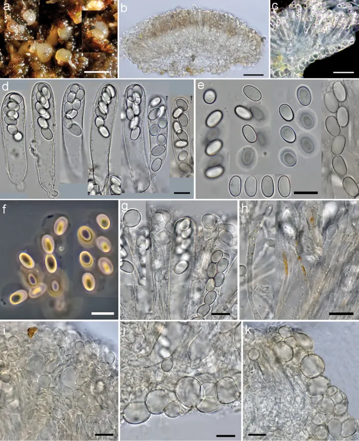
Coprotus epithecioides (CNF 2/10450, holotype). a Fresh apothecia on Rupicapra rupicapra dung b Cross section through the whole apothecia c Cross section in dark field d Asci e Freshly ejected ascospores glued together with a sheath and individual ascospores f Freshly ejected ascospores in phase contrast g Epithecioid paraphyses h Clavate paraphyses with pigment content i Epithecioid hymenial cover j Excipular flank k Marginal tissue. All elements observed in tap water and in living state, except two asci on d marked with a cross (†); Scale bars: a 0.5 mm, b, c 50 μm, d–k 10 μm, phot. N. Matočec & I. Kušan.
- Jenis
- Pelat ilmiah
- Kredit
- Ivana Kušan, Neven Matočec, Margita Jadan, Zdenko Tkalčec, Armin Mešić
- Lisensi
- CC BY 4.0
- Sumber media
- Buka entri sumber
Dokumentasi tambahan
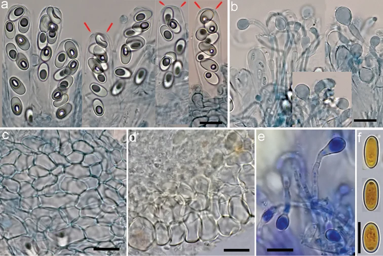
Coprotus epithecioides (CNF 2/10450, holotype). a Asci with ascospores containing de Bary bubbles, red markings show opercular delimitation b Paraphyses c Ectal excipulum from top view d Excipular flank f Paraphyses g Ascospores. a–c † CB d † MLZ e * CRB f † IKI. Scale bars: a–f 10 μm, phot. N. Matočec & I. Kušan.
Kredit dan penggunaan
Ivana Kušan, Neven Matočec, Margita Jadan, Zdenko Tkalčec, Armin Mešić
Ukuran dan format gambar disesuaikan tanpa pemotongan, retus, atau penambahan skala. Lisensi sumber dipertahankan.
Sumber profil
- [1] An overview of the genus Coprotus (Pezizales, Ascomycota) with notes on the type species and description of C. epithecioides sp. nov. — Coprotus epithecioides
- [2] Catalogue of Life Base2026-09-11 — Coprotus epithecioides
- [3] Dokumentasi visual — Coprotus epithecioides
- [4] Dokumentasi visual — Coprotus epithecioides
Rujukan halaman ini
Fungi. Coprotus epithecioides. Rilis data fungi-2026-10-12-expansion-2. https://fungi-atlas.mgadib.chatgpt.site/jamur/coprotus-epithecioides/.
Status: impor sumber; pemeriksaan editorial per profil belum selesai. Untuk klaim biologis, gunakan rujukan pada bagian terkait dan periksa konteks sumber aslinya.