Ciri dan struktur
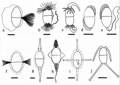
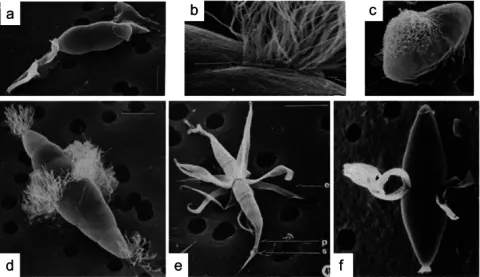
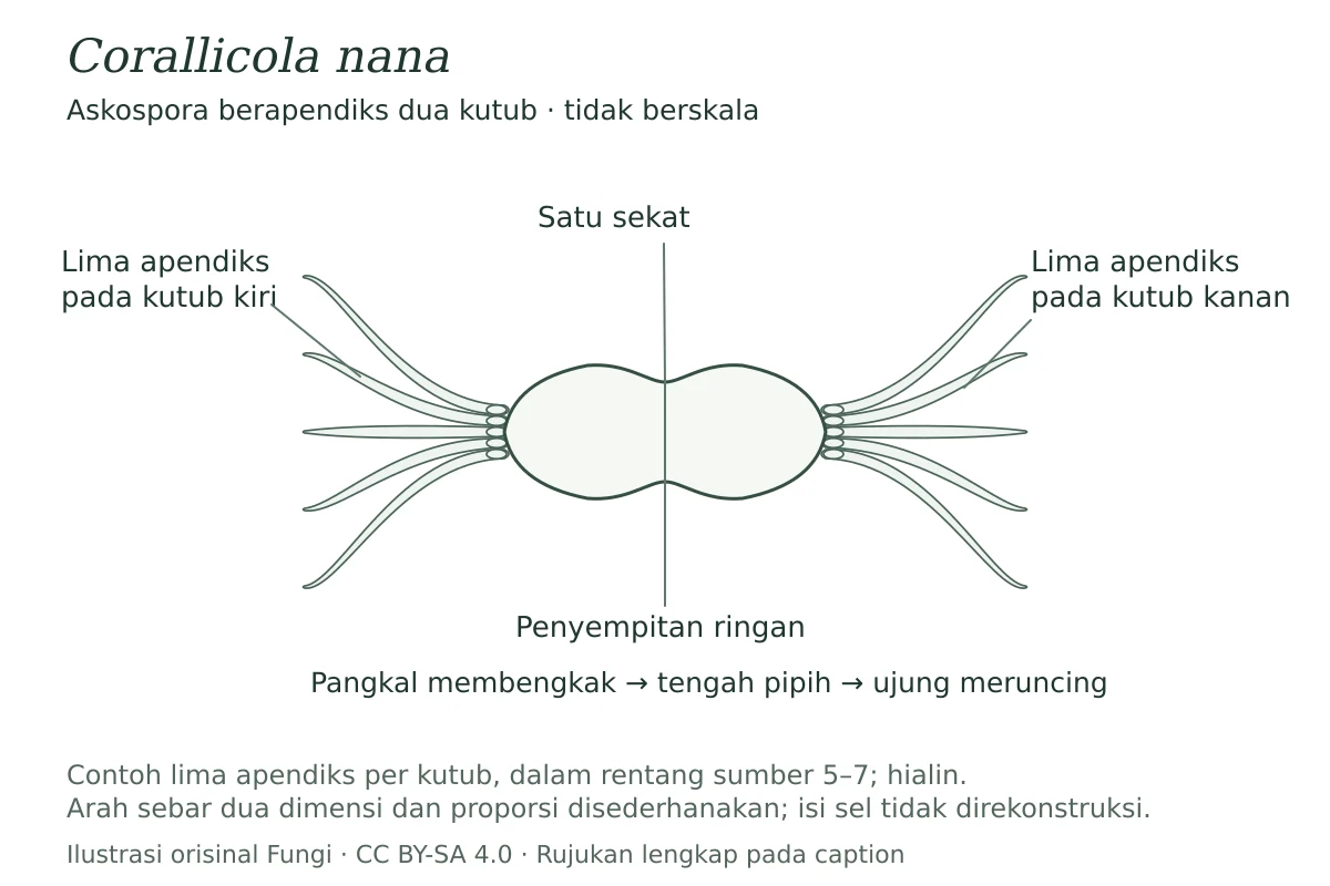
Saprobic on dead coral. Sexual morph. Ascomata are solitary or gregarious 80–95 µm in diam., subglobose, superficial, ostiolate, short papillate or epipapillate, subiculate, coriaceous, dark brown, sometimes covered by short brown hyphae. Peridium is 5–7 µm thick, composed of one or two layers of polygonal cells, forming a textura angularis, dark brown; at the base attached to the substrate with a thin subiculum that is more or less hyphoid (textura intricata) or dense (textura angularis). Pseudoparenchyma of thin-walled polygonal cells filling the centrum of young ascomata, deliquescing at ascospore maturity; no indication of pit-connections in the walls. Asci thin-walled, deliquescing before ascospore maturation. Ascospores range from 21–26.5 × 7–8.5 µm, ellipsoidal, 1-septate, slightly constricted at the septum, hyaline, appendaged at each end with 5–7 terminal appendages, 15–18 µm long, round and swollen at the base, 1.4–1.7 µm wide and flat in the middle, and tapering to the thin (0.5) µm round tip (Adapted from Volkmann-Kohlmeyer and Kohlmeyer 1992). Asexual morph. Undetermined.
Habitat dan peran ekologis
Saprobic on dead coral. Sexual morph. Ascomata are solitary or gregarious 80–95 µm in diam., subglobose, superficial, ostiolate, short papillate or epipapillate, subiculate, coriaceous, dark brown, sometimes covered by short brown hyphae. Peridium is 5–7 µm thick, composed of one or two layers of polygonal cells, forming a textura angularis, dark brown; at the base attached to the substrate with a thin subiculum that is more or less hyphoid (textura intricata) or dense (textura angularis). Pseudoparenchyma of thin-walled polygonal cells filling the centrum of young ascomata, deliquescing at ascospore maturity; no indication of pit-connections in the walls. Asci thin-walled, deliquescing before ascospore maturation. Ascospores range from 21–26.5 × 7–8.5 µm, ellipsoidal, 1-septate, slightly constricted at the septum, hyaline, appendaged at each end with 5–7 terminal appendages, 15–18 µm long, round and swollen at the base, 1.4–1.7 µm wide and flat in the middle, and tapering to the thin (0.5) µm round tip (Adapted from Volkmann-Kohlmeyer and Kohlmeyer 1992). Asexual morph. Undetermined.
- Habitat atau konteks temuan
- Informasi belum tersedia pada sumber rilis ini.
- Substrat
- Informasi belum tersedia pada sumber rilis ini.
- Peran atau konteks
- Pengurai
Reproduksi
Saprobic on dead coral. Sexual morph. Ascomata are solitary or gregarious 80–95 µm in diam., subglobose, superficial, ostiolate, short papillate or epipapillate, subiculate, coriaceous, dark brown, sometimes covered by short brown hyphae. Peridium is 5–7 µm thick, composed of one or two layers of polygonal cells, forming a textura angularis, dark brown; at the base attached to the substrate with a thin subiculum that is more or less hyphoid (textura intricata) or dense (textura angularis). Pseudoparenchyma of thin-walled polygonal cells filling the centrum of young ascomata, deliquescing at ascospore maturity; no indication of pit-connections in the walls. Asci thin-walled, deliquescing before ascospore maturation.
Catatan identitas dan cakupan
- Nama diterima mengikuti Catalogue of Life Base2026-09-11; uraian mempertahankan konsep dan nama sumber aslinya.
- Preserve original treatment localities, hosts and substrate scope. Materials examined describe documented specimens, not the full global range.
- Impor dan terjemahan otomatis belum diperiksa editorial per profil. Struktur reproduktif yang disebutkan tidak membuktikan seluruh siklus hidup diketahui.
- Pengukuran pada uraian mengikuti spesimen, kultur, dan metode sumber; angka tidak diperluas ke semua populasi.
Catatan distribusi dalam narasi mengikuti sumbernya. Dokumentasi spesimen atau foto mengikuti catatan sumber. Bukti wilayah Indonesia dan penilaian konservasi tidak disimpulkan dari gambar.
Melihat dengan konteks
Skema askospora Corallicola nana: elipsoidal, hialin, bersekat satu dengan penyempitan ringan, serta 5–7 apendiks di setiap kutub. Lima apendiks per kutub ditampilkan sebagai salah satu jumlah dalam rentang sumber; pangkal membengkak, bagian tengah pipih, dan ujung meruncing membulat. Tidak berskala; arah sebar apendiks disederhanakan dan isi sel tidak direkonstruksi.
- Jenis
- Ilustrasi skematis
- Kredit
- Ilustrasi orisinal Fungi berdasarkan uraian: Dayarathne MC, Jones EBG, Bahkali AH, Devadatha B, Hyde KD, Abdel-Wahab MA, Calabon MS, Correia P, Abdel-Aziz FA, Bakhit MS, Sakayaroj J, Prematunga C, Caeiro MF, Azevedo E, Asghari R, Apurillo CCS, Bhagya AT, Aumentado HD, Mukhopadhyay S, Phukhamsakda C, Chen Y, Maharachchikumbura SS, Guo SY, Pang KL. — Taxonomy and molecular evaluation of the Halosphaeriaceae (Microascales, Hypocreomycetidae) — Corallicola nana. Sumber: CC BY 4.0. Perubahan: skema anatomi konseptual, label dan caption Indonesia; bukan penyalinan gambar sumber.
- Lisensi
- CC BY-SA 4.0
- Sumber media
- Buka entri sumber
Sumber profil
- [1] Taxonomy and molecular evaluation of the Halosphaeriaceae (Microascales, Hypocreomycetidae) — Corallicola nana
- [2] Catalogue of Life Base2026-09-11 — Corallicola nana
Rujukan halaman ini
Fungi. Corallicola nana. Rilis data fungi-2026-10-12-expansion-2. https://fungi-atlas.mgadib.chatgpt.site/jamur/corallicola-nana/.
Status: impor sumber; pemeriksaan editorial per profil belum selesai. Untuk klaim biologis, gunakan rujukan pada bagian terkait dan periksa konteks sumber aslinya.