01 / Morfologi

Ciri dan struktur

Saprobic marine fungus. Sexual morph. Ascomata 138–590 µm, subglobose or ellipsoidal, immersed or partly immersed, ostiolate, papillate, subcarbonaceous, black, solitary or gregarious. Peridium 12–32 µm thick, composed of five to eight (to twenty) layers of irregular, polygonal, thick-walled cells, forming a textura angularis, merging into the pseudoparenchyma of the venter. Papillae or necks 35–320 µm long, 35–80(–140) µm in diam., conical or subcylindrical, centric or eccentric, dark, ostiolar canal periphysate. Asci 62.5–168(–200) × 20–48 µm, 8-spored, clavate, pedunculate, unitunicate, thin-walled, aphysoclastic. Ascospores 24–44.5 × 8–17 (–20) µm (excluding appendages), ellipsoidal or subfusiform, 1-septate, not or slightly constricted at the septum, hyaline, appendaged; at both ends with a small cap that may invert; around the septum three (rarely four) crescent-shaped, rigid, attenuate appendages, (12–)24.5–40 µm long, up to 1.3 µm in diam. at the base, obliquely attached to the septum, developing by fragmentation of the exospore. Asexual morph. Conidiophores absent or short, zero- to four-septate, simple, formed laterally on hyphae, hyaline to light brown or fuscous. Conidia (blastoconidia) (15–)20–34(–45) × (8–) 10–24 µm clavate, ovoid or obpyriform, two- to five-septate, constricted at the septa, straight or slightly curved, increasing in diameter from base to apex, formed singly on the conidiophores; apical cells subglobose, dark brown; basal cells conical or subcylindrical, subhyaline to light brown or fuscous. Chlamydospores are 6–16, catenulate µm, dark brown; chains up to 500 µm, long chains up to 35-celled, terminal, rarely intercalary, simple; cells increasing in diameter from base to apex, 7.5–19 µm long, globose or ellipsoidal (Description adapted from Kohlmeyer and Kohlmeyer 1979).

Sebagian uraian ditampilkan dalam bahasa sumber; terjemahannya belum tersedia atau belum lolos pemeriksaan.

Rujukan: [1]

Pengukuran mengikuti objek, spesimen atau kultur, dan metode yang disebutkan sumber. Baca uraian asli untuk konteks lengkap.

02 / Kehidupan

Habitat dan peran ekologis

Australia, Canada, Bulgaria, Denmark, France, Iceland, Germany, Japan, Liberia, Mexico, Portugal, Senegal, Spain, UK (England, Scotland, Wales), USA (California, Florida, Maryland, Washington).

Sebagian uraian ditampilkan dalam bahasa sumber; terjemahannya belum tersedia atau belum lolos pemeriksaan.

Rujukan: [1]

Habitat atau konteks temuan
Informasi belum tersedia pada sumber rilis ini.
Substrat
Informasi belum tersedia pada sumber rilis ini.
Peran atau konteks
Belum dikategorikan pada rilis ini.
Temukan hubungan serupa
03 / Siklus hidup

Reproduksi

Saprobic marine fungus. Sexual morph. Ascomata 138–590 µm, subglobose or ellipsoidal, immersed or partly immersed, ostiolate, papillate, subcarbonaceous, black, solitary or gregarious. Peridium 12–32 µm thick, composed of five to eight (to twenty) layers of irregular, polygonal, thick-walled cells, forming a textura angularis, merging into the pseudoparenchyma of the venter. Papillae or necks 35–320 µm long, 35–80(–140) µm in diam., conical or subcylindrical, centric or eccentric, dark, ostiolar canal periphysate. Asci 62.5–168(–200) × 20–48 µm, 8-spored, clavate, pedunculate, unitunicate, thin-walled, aphysoclastic. Ascospores 24–44.5 × 8–17 (–20) µm (excluding appendages), ellipsoidal or subfusiform, 1-septate, not or slightly constricted at the septum, hyaline, appendaged; at both ends with a small cap that may invert; around the septum three (rarely four) crescent-shaped, rigid, attenuate appendages, (12–)24.5–40 µm long, up to 1.3 µm in diam. at the base, obliquely attached to the septum, developing by fragmentation of the exospore.

Sebagian uraian ditampilkan dalam bahasa sumber; terjemahannya belum tersedia atau belum lolos pemeriksaan.

Rujukan: [1]

Sinonim menurut acuan taksonomi rilis: Culcitalna achraspora; Halosphaeria mediosetigera; Halosphaeria mediosetigera var. grandispora; Trichocladium achrasporum
04 / Batas informasi

Catatan identitas dan cakupan

  • Nama diterima mengikuti Catalogue of Life Base2026-09-11; uraian mempertahankan konsep dan nama sumber aslinya.
  • Preserve original treatment localities, hosts and substrate scope. Materials examined describe documented specimens, not the full global range.
  • Impor dan terjemahan otomatis belum diperiksa editorial per profil. Struktur reproduktif yang disebutkan tidak membuktikan seluruh siklus hidup diketahui.
  • Pengukuran pada uraian mengikuti spesimen, kultur, dan metode sumber; angka tidak diperluas ke semua populasi.

Catatan distribusi dalam narasi mengikuti sumbernya. Dokumentasi spesimen atau foto mengikuti catatan sumber. Bukti wilayah Indonesia dan penilaian konservasi tidak disimpulkan dari gambar.

05 / Dokumentasi

Melihat dengan konteks

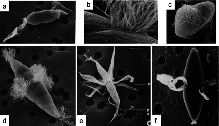
Scanning electron micrographs of different types of ascospore appendages in the Halosphaeriaceae. a. Remispora maritima with polar appendages; b. Nereiospora cristata hair-like equatorial appendages arising from a pad (arrowed); c. Nimbospora effusa with sticky appendage; d. Nereiospora comata with hair-like polar and equatorial appendages; e. Corollospora intermedia with polar spine peritrichous appendages apically and at the central septum (e=equatorial appendages, p=polar spine appendage, s=polar secondary appendages); f. Halosphaeriopsis mediosetigera with polar caps and lunate equatorial appendages. Scale bars: 10 mm (a, c–f); 1 mm (b).

Jenis
Pelat ilmiah
Kredit
Monika C. Dayarathne, E. B. Gareth Jones, Ali H. Bahkali, Bandarupalli Devadatha, Kevin D. Hyde, Mohamed A. Abdel-Wahab, Mark S. Calabon, Pedro Correia, Faten A. Abdel-Aziz, Mahmoud S. Bakhit, Jariya Sakayaroj, Chasika Prematunga, Maria F. Caeiro, Egídia Azevedo, Raheleh Asghari, Carlo Chris S. Apurillo, Amuhenage Tharindu Bhagya, Herbert Dustin Aumentado, Samhita Mukhopadhyay, Chayanard Phukhamsakda, Yanpeng Chen, Sajeewa S. Maharachchikumbura, Sheng-Yu Guo, Ka-Lai Pang
Lisensi
CC BY 4.0
Sumber media
Buka entri sumber

Gambar menunjukkan objek menurut identifikasi sumber, bukan verifikasi independen spesimen oleh Fungi. Tidak ada skala baru yang ditambahkan.

06 / Rujukan

Sumber profil

  1. [1] Taxonomy and molecular evaluation of the Halosphaeriaceae (Microascales, Hypocreomycetidae) — Halosphaeriopsis mediosetigeraBagian sumber: PMC13329432_section_289
    Tanggal penghimpunan sumber: 2026-10-12
    Teks sumber: CC BY 4.0 · Dayarathne MC, Jones EBG, Bahkali AH, Devadatha B, Hyde KD, Abdel-Wahab MA, Calabon MS, Correia P, Abdel-Aziz FA, Bakhit MS, Sakayaroj J, Prematunga C, Caeiro MF, Azevedo E, Asghari R, Apurillo CCS, Bhagya AT, Aumentado HD, Mukhopadhyay S, Phukhamsakda C, Chen Y, Maharachchikumbura SS, Guo SY, Pang KL.
  2. [2] Catalogue of Life Base2026-09-11 — Halosphaeriopsis mediosetigeraBagian sumber: ColDP NameUsage.tsv ID 3JG65; accepted species; source 2073
    Tanggal penghimpunan sumber: 2026-10-12
  3. [3] Dokumentasi visual — Halosphaeriopsis mediosetigeraBagian sumber: Individual media record; identification follows the source, not independent determination
    Tanggal penghimpunan sumber: 2026-10-12
    Media sumber: CC BY 4.0 · Monika C. Dayarathne, E. B. Gareth Jones, Ali H. Bahkali, Bandarupalli Devadatha, Kevin D. Hyde, Mohamed A. Abdel-Wahab, Mark S. Calabon, Pedro Correia, Faten A. Abdel-Aziz, Mahmoud S. Bakhit, Jariya Sakayaroj, Chasika Prematunga, Maria F. Caeiro, Egídia Azevedo, Raheleh Asghari, Carlo Chris S. Apurillo, Amuhenage Tharindu Bhagya, Herbert Dustin Aumentado, Samhita Mukhopadhyay, Chayanard Phukhamsakda, Yanpeng Chen, Sajeewa S. Maharachchikumbura, Sheng-Yu Guo, Ka-Lai Pang

Adaptasi teks: CC BY 4.0. Dayarathne MC, Jones EBG, Bahkali AH, Devadatha B, Hyde KD, Abdel-Wahab MA, Calabon MS, Correia P, Abdel-Aziz FA, Bakhit MS, Sakayaroj J, Prematunga C, Caeiro MF, Azevedo E, Asghari R, Apurillo CCS, Bhagya AT, Aumentado HD, Mukhopadhyay S, Phukhamsakda C, Chen Y, Maharachchikumbura SS, Guo SY, Pang KL. — Taxonomy and molecular evaluation of the Halosphaeriaceae (Microascales, Hypocreomycetidae) — Halosphaeriopsis mediosetigera

Uraian asli dan riwayat sumber

Riwayat atau halaman sumber

Ringkasan sumber

Saprobic marine fungus. Sexual morph. Ascomata 138–590 µm, subglobose or ellipsoidal, immersed or partly immersed, ostiolate, papillate, subcarbonaceous, black, solitary or gregarious. Peridium 12–32 µm thick, composed of five to eight (to twenty) layers of irregular, polygonal, thick-walled cells, forming a textura angularis, merging into the pseudoparenchyma of the venter. Papillae or necks 35–320 µm long, 35–80(–140) µm in diam., conical or subcylindrical, centric or eccentric, dark, ostiolar canal periphysate.

Revisi sumber

Morfologi sumber

Saprobic marine fungus. Sexual morph. Ascomata 138–590 µm, subglobose or ellipsoidal, immersed or partly immersed, ostiolate, papillate, subcarbonaceous, black, solitary or gregarious. Peridium 12–32 µm thick, composed of five to eight (to twenty) layers of irregular, polygonal, thick-walled cells, forming a textura angularis, merging into the pseudoparenchyma of the venter. Papillae or necks 35–320 µm long, 35–80(–140) µm in diam., conical or subcylindrical, centric or eccentric, dark, ostiolar canal periphysate. Asci 62.5–168(–200) × 20–48 µm, 8-spored, clavate, pedunculate, unitunicate, thin-walled, aphysoclastic. Ascospores 24–44.5 × 8–17 (–20) µm (excluding appendages), ellipsoidal or subfusiform, 1-septate, not or slightly constricted at the septum, hyaline, appendaged; at both ends with a small cap that may invert; around the septum three (rarely four) crescent-shaped, rigid, attenuate appendages, (12–)24.5–40 µm long, up to 1.3 µm in diam. at the base, obliquely attached to the septum, developing by fragmentation of the exospore. Asexual morph. Conidiophores absent or short, zero- to four-septate, simple, formed laterally on hyphae, hyaline to light brown or fuscous. Conidia (blastoconidia) (15–)20–34(–45) × (8–) 10–24 µm clavate, ovoid or obpyriform, two- to five-septate, constricted at the septa, straight or slightly curved, increasing in diameter from base to apex, formed singly on the conidiophores; apical cells subglobose, dark brown; basal cells conical or subcylindrical, subhyaline to light brown or fuscous. Chlamydospores are 6–16, catenulate µm, dark brown; chains up to 500 µm, long chains up to 35-celled, terminal, rarely intercalary, simple; cells increasing in diameter from base to apex, 7.5–19 µm long, globose or ellipsoidal (Description adapted from Kohlmeyer and Kohlmeyer 1979).

Revisi sumber

Ekologi sumber

Australia, Canada, Bulgaria, Denmark, France, Iceland, Germany, Japan, Liberia, Mexico, Portugal, Senegal, Spain, UK (England, Scotland, Wales), USA (California, Florida, Maryland, Washington).

Revisi sumber

Struktur reproduksi sumber

Saprobic marine fungus. Sexual morph. Ascomata 138–590 µm, subglobose or ellipsoidal, immersed or partly immersed, ostiolate, papillate, subcarbonaceous, black, solitary or gregarious. Peridium 12–32 µm thick, composed of five to eight (to twenty) layers of irregular, polygonal, thick-walled cells, forming a textura angularis, merging into the pseudoparenchyma of the venter. Papillae or necks 35–320 µm long, 35–80(–140) µm in diam., conical or subcylindrical, centric or eccentric, dark, ostiolar canal periphysate. Asci 62.5–168(–200) × 20–48 µm, 8-spored, clavate, pedunculate, unitunicate, thin-walled, aphysoclastic. Ascospores 24–44.5 × 8–17 (–20) µm (excluding appendages), ellipsoidal or subfusiform, 1-septate, not or slightly constricted at the septum, hyaline, appendaged; at both ends with a small cap that may invert; around the septum three (rarely four) crescent-shaped, rigid, attenuate appendages, (12–)24.5–40 µm long, up to 1.3 µm in diam. at the base, obliquely attached to the septum, developing by fragmentation of the exospore.

Revisi sumber

Rujukan halaman ini

Fungi. Halosphaeriopsis mediosetigera. Rilis data fungi-2026-10-12-expansion-2. https://fungi-atlas.mgadib.chatgpt.site/jamur/halosphaeriopsis-mediosetigera/.

Status: impor sumber; pemeriksaan editorial per profil belum selesai. Untuk klaim biologis, gunakan rujukan pada bagian terkait dan periksa konteks sumber aslinya.