Ciri dan struktur
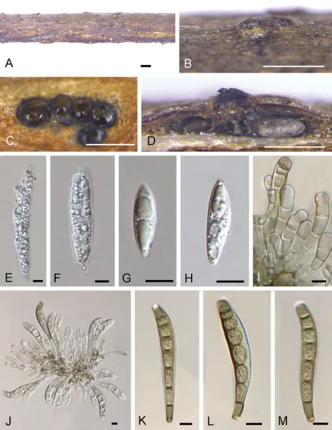
Associated with canker on twigs of Hevea brasiliensis. Asexual morph: Conidiomata acervular, solitary, erumpent through the outer periderm layers of host, scattered, surface tissues above slightly dome-shaped, black, velvety, formed of brown cell, thick-walled textura angularis, 145–540 µm diam. Conidiophores short, cylindrical, apically pale brown, paler at the base, smooth, septate, branched at the base, arising from basal stroma, 22–37 × 4–8 μm (x ‒ = 28.5 × 5.6 μm, n = 15). Conidiogenous cell annellidic, integrated, terminal, cylindrical, medium brown, truncate apex, with 1-3 slightly percurrent proliferations, 6–17 µm long (x ‒ = 10.7 μm, n = 20). Conidia curved, clavate to fusiform, dark brown, smooth-walled, 4–6-pseudo-septa, sometimes with apical and basal cells darker than other cells, rounded or sometime truncate at apex, truncate and black at the base, (40–)43–53(–68) × (14–)15–20 μm (x ‒ = 48.7 × 17.3 μm, n = 85). Appressoria hyaline, globose to sub globose, thick-walled, 4–11 μm wide (x ‒ = 7.1 μm, n = 20). Conidia germinated on MEA within 24 h with germ tubes produced from one or both end cells, mostly from basal cell of conidia. Colonies on MEA reaching 20–25 mm diam. after 4 weeks at 25–30 °C, colonies circular, medium dense, cottony, margin wavy, superficial, slightly effuse, radially striated; colony from above, white, edges with more aerial mycelium than centre in the beginning and later become white grey, smooth with edge entire; from below: white to cream at the margin, yellowish-green in the centre in the beginning and later become dark green; not producing pigmentation in agar.
Habitat dan peran ekologis
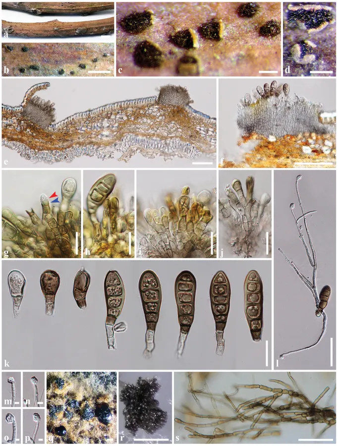
Conidia germinated on MEA within 24 h with germ tubes produced from one or both end cells, mostly from basal cell of conidia. Colonies on MEA reaching 20–25 mm diam. after 4 weeks at 25–30 °C, colonies circular, medium dense, cottony, margin wavy, superficial, slightly effuse, radially striated; colony from above, white, edges with more aerial mycelium than centre in the beginning and later become white grey, smooth with edge entire; from below: white to cream at the margin, yellowish-green in the centre in the beginning and later become dark green; not producing pigmentation in agar. Colonies on PDA reaching 10–15 mm diam. after 4 weeks at 25–30 °C, colonies circular, medium dense, cottony, slightly effuse, dark green with brown aerial mycelium on surface; not producing pigmentation in agar. Conidial masses were observed in PDA culture after 6 months at 25–30 °C. Mass of conidia dark brown to black, extruding on colony or tip of mycelium (Figure 2 q, r). Mycelium superficial and immersed, dark brown, hyphae branched, septate, constricted at septa, thick, smooth-walled (Figure 2 s).
- Habitat atau konteks temuan
- kultur yang didokumentasikan
- Substrat
- Informasi belum tersedia pada sumber rilis ini.
- Peran atau konteks
- Belum dikategorikan pada rilis ini.
Reproduksi
Associated with canker on twigs of Hevea brasiliensis. Asexual morph: Conidiomata acervular, solitary, erumpent through the outer periderm layers of host, scattered, surface tissues above slightly dome-shaped, black, velvety, formed of brown cell, thick-walled textura angularis, 145–540 µm diam. Conidiophores short, cylindrical, apically pale brown, paler at the base, smooth, septate, branched at the base, arising from basal stroma, 22–37 × 4–8 μm (x ‒ = 28.5 × 5.6 μm, n = 15). Conidiogenous cell annellidic, integrated, terminal, cylindrical, medium brown, truncate apex, with 1-3 slightly percurrent proliferations, 6–17 µm long (x ‒ = 10.7 μm, n = 20). Conidia curved, clavate to fusiform, dark brown, smooth-walled, 4–6-pseudo-septa, sometimes with apical and basal cells darker than other cells, rounded or sometime truncate at apex, truncate and black at the base, (40–)43–53(–68) × (14–)15–20 μm (x ‒ = 48.7 × 17.3 μm, n = 85).
Catatan identitas dan cakupan
- Nama diterima mengikuti Catalogue of Life Base2026-09-11; uraian mempertahankan konsep dan nama sumber aslinya.
- Preserve original treatment localities, hosts and substrate scope. Materials examined describe documented specimens, not the full global range.
- Impor dan terjemahan otomatis belum diperiksa editorial per profil. Struktur reproduktif yang disebutkan tidak membuktikan seluruh siklus hidup diketahui.
- Pengukuran pada uraian mengikuti spesimen, kultur, dan metode sumber; angka tidak diperluas ke semua populasi.
Catatan distribusi dalam narasi mengikuti sumbernya. Dokumentasi spesimen atau foto mengikuti catatan sumber. Bukti wilayah Indonesia dan penilaian konservasi tidak disimpulkan dari gambar.
Melihat dengan konteks
Coryneum heveanum (MFLU 18-0936). a−d Conidiomata on host surface e−f Acervuli g Conidiogenesis (annellidic; red arrow, proliferation; blue arrow) h−j Conidiophores, conidiogeneous cells with conidia k Conidia l Germinated spores m−p Appressoria q−r Mass of conidia on PDA after 6 months s Mycelium on PDA after 6 months. Scale bars: 5 mm (a), 1000 µm (b), 200 µm (c, d, r), 100 µm (e, f, q), 20 µm (g−k), 50 µm (l, s), 5 µm (m−p).
- Jenis
- Pelat ilmiah
- Kredit
- Chanokned Senwanna, Kevin D. Hyde, Rungtiwa Phookamsak, E. B. Gareth Jones, Ratchadawan Cheewangkoon
- Lisensi
- CC BY 4.0
- Sumber media
- Buka entri sumber
Sumber profil
- [1] Coryneum heveanum sp. nov. (Coryneaceae, Diaporthales) on twigs of Para rubber in Thailand — Coryneum heveanum
- [2] Catalogue of Life Base2026-09-11 — Coryneum heveanum
- [3] Dokumentasi visual — Coryneum heveanum
Rujukan halaman ini
Fungi. Coryneum heveanum. Rilis data fungi-2026-10-12-expansion-2. https://fungi-atlas.mgadib.chatgpt.site/jamur/coryneum-heveanum/.
Status: impor sumber; pemeriksaan editorial per profil belum selesai. Untuk klaim biologis, gunakan rujukan pada bagian terkait dan periksa konteks sumber aslinya.