Ciri dan struktur
Sexual morph: not observed. Asexual morph: Pycnidial stromata pulvinate, immersed in bark, scattered, erumpent through the surface of bark. Conceptacle absent. Ostiole dark grey to black, conspicuous, at the same level as the disc, (50–)65–75(–82) μm diam. Locules undivided, circular to ovoid, (430–)550–720(–810) µm diam. Peridium comprising few layers of cells of textura angularis, with innermost layer brown, outer layer brown to dark brown. Conidiophores hyaline, unbranched, thin-walled, filamentous. Conidiogenous cells enteroblastic polyphialidic, (6.5–)8.4–12.5(–15.3) × 0.9–1.4 μm (x̄ = 10.2 × 1.2 μm). Conidia hyaline, allantoid, smooth, aseptate, thin-walled, (3.2–)3.4–5.4(–6.2) × 1–1.5 μm (x̄ = 4.7 × 1.3 μm).
Habitat dan peran ekologis
On PDA at 25 °C in darkness. Cultures are initially white, becoming olivaceous buff in centre after 7 d and finally olivaceous at 30 d. The colony is flat, thin with a felt and tight texture in centre. Pycnidia distributed irregularly on medium surface.
- Habitat atau konteks temuan
- kultur yang didokumentasikan
- Substrat
- Informasi belum tersedia pada sumber rilis ini.
- Peran atau konteks
- Belum dikategorikan pada rilis ini.
Reproduksi
Sexual morph: not observed. Asexual morph: Pycnidial stromata pulvinate, immersed in bark, scattered, erumpent through the surface of bark. Conceptacle absent. Ostiole dark grey to black, conspicuous, at the same level as the disc, (50–)65–75(–82) μm diam. Locules undivided, circular to ovoid, (430–)550–720(–810) µm diam. Peridium comprising few layers of cells of textura angularis, with innermost layer brown, outer layer brown to dark brown. Conidiophores hyaline, unbranched, thin-walled, filamentous. Conidiogenous cells enteroblastic polyphialidic, (6.5–)8.4–12.5(–15.3) × 0.9–1.4 μm (x̄ = 10.2 × 1.2 μm). Conidia hyaline, allantoid, smooth, aseptate, thin-walled, (3.2–)3.4–5.4(–6.2) × 1–1.5 μm (x̄ = 4.7 × 1.3 μm).
Catatan identitas dan cakupan
- Nama diterima mengikuti Catalogue of Life Base2026-09-11; uraian mempertahankan konsep dan nama sumber aslinya.
- Preserve original treatment localities, hosts and substrate scope. Materials examined describe documented specimens, not the full global range.
- Impor dan terjemahan otomatis belum diperiksa editorial per profil. Struktur reproduktif yang disebutkan tidak membuktikan seluruh siklus hidup diketahui.
- Pengukuran pada uraian mengikuti spesimen, kultur, dan metode sumber; angka tidak diperluas ke semua populasi.
Catatan distribusi dalam narasi mengikuti sumbernya. Dokumentasi spesimen atau foto mengikuti catatan sumber. Bukti wilayah Indonesia dan penilaian konservasi tidak disimpulkan dari gambar.
Melihat dengan konteks
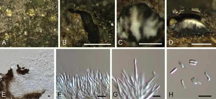
Cytospora myrtagena on Castanea mollissima (BJFC -S1704). A, B Habit of conidiomata on branches C, E transverse section of conidiomata D longitudinal section through conidiomata F, G conidiogenous cells attached with conidia H conidia. Scale bars: 500 μm (B–D), 5 μm (E, G), 10 μm (H).
- Jenis
- Pelat ilmiah
- Kredit
- Ning Jiang, Qin Yang, Xin-Lei Fan, Cheng-Ming Tian
- Lisensi
- CC BY 4.0
- Sumber media
- Buka entri sumber
Sumber profil
- [1] Identification of six Cytospora species on Chinese chestnut in China — Cytospora myrtagena
- [2] Catalogue of Life Base2026-09-11 — Cytospora myrtagena
- [3] Dokumentasi visual — Cytospora myrtagena
Rujukan halaman ini
Fungi. Cytospora myrtagena. Rilis data fungi-2026-10-12-expansion-2. https://fungi-atlas.mgadib.chatgpt.site/jamur/cytospora-myrtagena/.
Status: impor sumber; pemeriksaan editorial per profil belum selesai. Untuk klaim biologis, gunakan rujukan pada bagian terkait dan periksa konteks sumber aslinya.