Ciri dan struktur
Characterised by larger J-shaped β-conidia. Colonies on PDA attaining 9 cm diam. after 10 days; coralloid with feathery branches at margin, adpressed, with apparent aerial mycelium, with numerous irregularly zonate dark stromata, isabelline becoming lighter towards the margin; reverse similar to surface, with zonations. Conidiomata pycnidial, multilocular, scattered, abundant on PDA after 3 wks, subglobose to irregular, 1.5–1.8 mm diam., ostiolate, with up to 1 mm necks when present. Conidiophores formed from the inner layer of the locular wall, sometimes reduced to conidiogenous cells, when present 1-septate, hyaline to pale yellowish-brown, cylindrical, 10–23 × 1–2.5 μm. Conidiogenous cells cylindrical to flexuous, tapered towards apex, hyaline, 8–18 × 1.5–3 μm. Alpha conidia abundant, fusiform, narrowed towards apex and base, mostly biguttulate, hyaline, 4.5–9 × 2–3.5 μm. Beta conidia scarce to abundant, flexuous to J-shaped, hyaline, 17.5–32 × 1–2 μm. Perithecia not seen.
Habitat dan peran ekologis
Colonies on PDA attaining 9 cm diam. after 10 days; coralloid with feathery branches at margin, adpressed, with apparent aerial mycelium, with numerous irregularly zonate dark stromata, isabelline becoming lighter towards the margin; reverse similar to surface, with zonations. Conidiomata pycnidial, multilocular, scattered, abundant on PDA after 3 wks, subglobose to irregular, 1.5–1.8 mm diam., ostiolate, with up to 1 mm necks when present. Conidiophores formed from the inner layer of the locular wall, sometimes reduced to conidiogenous cells, when present 1-septate, hyaline to pale yellowish-brown, cylindrical, 10–23 × 1–2.5 μm. Conidiogenous cells cylindrical to flexuous, tapered towards apex, hyaline, 8–18 × 1.5–3 μm. Alpha conidia abundant, fusiform, narrowed towards apex and base, mostly biguttulate, hyaline, 4.5–9 × 2–3.5 μm. Beta conidia scarce to abundant, flexuous to J-shaped, hyaline, 17.5–32 × 1–2 μm. Perithecia not seen. Isolated from leaves of Millettia reticulata in China
- Habitat atau konteks temuan
- kultur yang didokumentasikan
- Substrat
- Informasi belum tersedia pada sumber rilis ini.
- Peran atau konteks
- Belum dikategorikan pada rilis ini.
Reproduksi
Characterised by larger J-shaped β-conidia. Colonies on PDA attaining 9 cm diam. after 10 days; coralloid with feathery branches at margin, adpressed, with apparent aerial mycelium, with numerous irregularly zonate dark stromata, isabelline becoming lighter towards the margin; reverse similar to surface, with zonations. Conidiomata pycnidial, multilocular, scattered, abundant on PDA after 3 wks, subglobose to irregular, 1.5–1.8 mm diam., ostiolate, with up to 1 mm necks when present. Conidiophores formed from the inner layer of the locular wall, sometimes reduced to conidiogenous cells, when present 1-septate, hyaline to pale yellowish-brown, cylindrical, 10–23 × 1–2.5 μm. Conidiogenous cells cylindrical to flexuous, tapered towards apex, hyaline, 8–18 × 1.5–3 μm.
Catatan identitas dan cakupan
- Nama diterima mengikuti Catalogue of Life Base2026-09-11; uraian mempertahankan konsep dan nama sumber aslinya.
- Preserve original treatment localities, hosts and substrate scope. Materials examined describe documented specimens, not the full global range.
- Impor dan terjemahan otomatis belum diperiksa editorial per profil. Struktur reproduktif yang disebutkan tidak membuktikan seluruh siklus hidup diketahui.
- Pengukuran pada uraian mengikuti spesimen, kultur, dan metode sumber; angka tidak diperluas ke semua populasi.
Catatan distribusi dalam narasi mengikuti sumbernya. Dokumentasi spesimen atau foto mengikuti catatan sumber. Bukti wilayah Indonesia dan penilaian konservasi tidak disimpulkan dari gambar.
Melihat dengan konteks
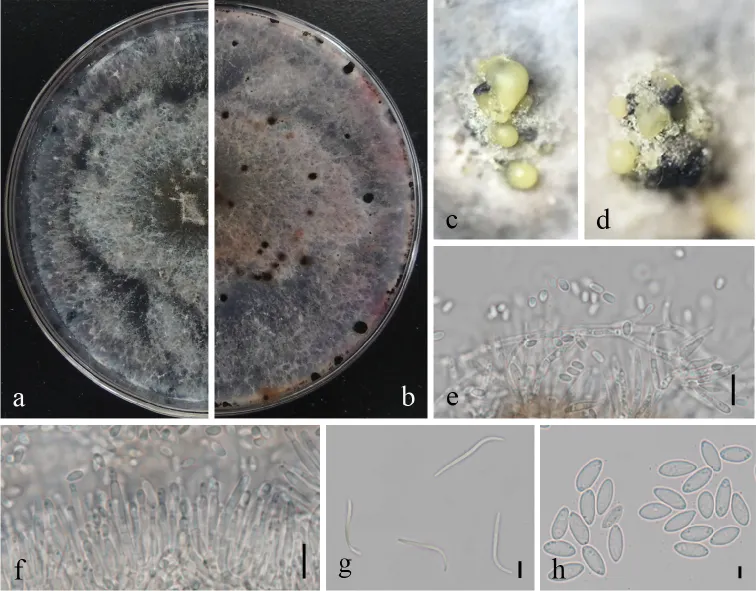
Diaporthe millettiae (GUCC9167). a–b upper (a) and lower (b) surface of colony on PDA c–d conidiomata e–f conidiophores, conidiogenous loci and conidia g β-conidia h α-conidia. Scale bars: 20 µm (e, f), 10 µm (g, h).
- Jenis
- Pelat ilmiah
- Kredit
- Hui Long, Qian Zhang, Yuan-Yuan Hao, Xian-Qiang Shao, Xiao-Xing Wei, Kevin D. Hyde, Yong Wang, De-Gang Zhao
- Lisensi
- CC BY 4.0
- Sumber media
- Buka entri sumber
Sumber profil
- [1] Diaporthe species in south-western China — Diaporthe millettiae
- [2] Catalogue of Life Base2026-09-11 — Diaporthe millettiae
- [3] Dokumentasi visual — Diaporthe millettiae
Rujukan halaman ini
Fungi. Diaporthe millettiae. Rilis data fungi-2026-10-12-expansion-2. https://fungi-atlas.mgadib.chatgpt.site/jamur/diaporthe-millettiae/.
Status: impor sumber; pemeriksaan editorial per profil belum selesai. Untuk klaim biologis, gunakan rujukan pada bagian terkait dan periksa konteks sumber aslinya.