Ciri dan struktur
Saprobic on decaying branches of Camellia oleifera. Sexual morph: Not observed. Asexual morph: Coelomycetous, Conidiomata 330–394 × 215–230 μm (x̄ = 362 × 223 μm, n = 10), immersed, erumpent, gregarious, dark brown to black, subglobose, unilocular. Ostiole 48.5–67 μm diam., central. Peridium 29–38 μm wide, thick-walled, outer and inner layers composed of 1–2 layers dark brown textura angularis, intermediate layers composed of 3–5 layers of hyaline cells of textura angularis. Conidiophores reduced to conidiogenous cells. Conidiogenous cells 8.5–12 × 3–5 μm (x̄ = 10 × 4 μm, n = 20), cylindrical, thin-walled, hyaline, holoblastic, indeterminate, proliferating at the same level to produce periclinal thickenings, or proliferating percurrently giving rise to 2–3 indistinct annellations. Conidia 19–21 × 9.5–11 μm (x̄ = 20 × 10.5 μm, n = 30), L/W ratio = 2, oblong, with broadly rounded apex and truncate base, thick-walled, wall externally smooth, roughened on the inner surface, hyaline, aseptate, becoming dark brown when aged. Colonies on PDA initially olivaceous buff in the center of the colony and white at the edge, becoming olivaceous within 7 d on the surface, with smooth edge.
Habitat dan peran ekologis
Saprobic on decaying branches of Camellia oleifera. Sexual morph: Not observed. Asexual morph: Coelomycetous, Conidiomata 330–394 × 215–230 μm (x̄ = 362 × 223 μm, n = 10), immersed, erumpent, gregarious, dark brown to black, subglobose, unilocular. Ostiole 48.5–67 μm diam., central. Peridium 29–38 μm wide, thick-walled, outer and inner layers composed of 1–2 layers dark brown textura angularis, intermediate layers composed of 3–5 layers of hyaline cells of textura angularis. Conidiophores reduced to conidiogenous cells. Conidiogenous cells 8.5–12 × 3–5 μm (x̄ = 10 × 4 μm, n = 20), cylindrical, thin-walled, hyaline, holoblastic, indeterminate, proliferating at the same level to produce periclinal thickenings, or proliferating percurrently giving rise to 2–3 indistinct annellations. Conidia 19–21 × 9.5–11 μm (x̄ = 20 × 10.5 μm, n = 30), L/W ratio = 2, oblong, with broadly rounded apex and truncate base, thick-walled, wall externally smooth, roughened on the inner surface, hyaline, aseptate, becoming dark brown when aged. Colonies on PDA initially olivaceous buff in the center of the colony and white at the edge, becoming olivaceous within 7 d on the surface, with smooth edge. China, Sichuan Province, Jiangyou City, Shuanghe County, 31°54'10"N, 104°55'57"E, elevation 657 m, on dead branches of Camellia oleifera, 11 th July 2021, W.L Li, 286 (HUEST 22.0069), living culture UESTCC 22.0068; ibid., 289 (HUEST 22.0068), living culture UESTCC 22.0067; ibid., Guangyuan city, Qingchuan County, 32°40'38"N, 105°28'57"E, elevation 634 m, on dead branches of Olea europaea, 20 th April 2021, W.L Li, 188 (HUEST 22.0065), living culture UESTCC 22.0064; ibid., 257 (HUEST 22.0070), living culture UESTCC 22.0069; ibid., on dead branches of Vernicia fordii, 20 th April 2021, W.L Li, 238 (HUEST 22.0066), living culture UESTCC 22.0065; ibid., Chengdu City, Pidu District, 30°49'27"N, 103°47'42"E, elevation 442 m, on dead branches of Pistacia chinensis, 5 th March 2021, W.L Li, A61 (HUEST 22.0064), living culture UESTCC 22.0063.
- Habitat atau konteks temuan
- kultur yang didokumentasikan
- Substrat
- kayu
- Peran atau konteks
- Pengurai
Reproduksi
Saprobic on decaying branches of Camellia oleifera. Sexual morph: Not observed. Asexual morph: Coelomycetous, Conidiomata 330–394 × 215–230 μm (x̄ = 362 × 223 μm, n = 10), immersed, erumpent, gregarious, dark brown to black, subglobose, unilocular. Ostiole 48.5–67 μm diam., central. Peridium 29–38 μm wide, thick-walled, outer and inner layers composed of 1–2 layers dark brown textura angularis, intermediate layers composed of 3–5 layers of hyaline cells of textura angularis. Conidiophores reduced to conidiogenous cells. Conidiogenous cells 8.5–12 × 3–5 μm (x̄ = 10 × 4 μm, n = 20), cylindrical, thin-walled, hyaline, holoblastic, indeterminate, proliferating at the same level to produce periclinal thickenings, or proliferating percurrently giving rise to 2–3 indistinct annellations.
Catatan identitas dan cakupan
- Nama diterima mengikuti Catalogue of Life Base2026-09-11; uraian mempertahankan konsep dan nama sumber aslinya.
- Preserve original treatment localities, hosts and substrate scope. Materials examined describe documented specimens, not the full global range.
- Impor dan terjemahan otomatis belum diperiksa editorial per profil. Struktur reproduktif yang disebutkan tidak membuktikan seluruh siklus hidup diketahui.
- Pengukuran pada uraian mengikuti spesimen, kultur, dan metode sumber; angka tidak diperluas ke semua populasi.
Catatan distribusi dalam narasi mengikuti sumbernya. Dokumentasi spesimen atau foto mengikuti catatan sumber. Bukti wilayah Indonesia dan penilaian konservasi tidak disimpulkan dari gambar.
Melihat dengan konteks
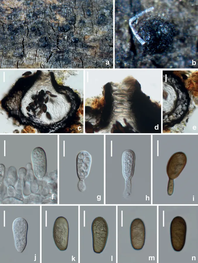
Diplodia mutila (HUEST 22.0069, new host record) a, b appearance of conidiomata on natural substrate c vertical section of conidioma d ostiole e section of peridium f–i conidiogenous cells and developing conidia j hyaline immature conidium k–n mature brown conidia. Scale bars: 40 μm (c); 20 μm (d, e); 10 μm (f–n).
- Jenis
- Pelat ilmiah
- Kredit
- Wen-Li Li, Rui-Ru Liang, Asha J. Dissanayake, Jian-Kui Liu
- Lisensi
- CC BY 4.0
- Sumber media
- Buka entri sumber
Dokumentasi tambahan
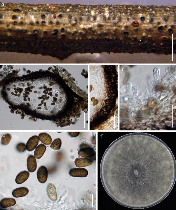
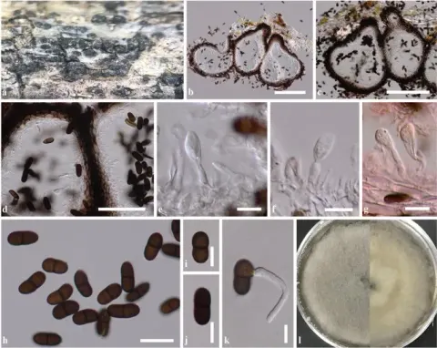
Diplodia mutila (MFLU 18-0912). a Appearance of conidiomata on the host. b Section through the conidioma. c Pycnidial wall. d Immature conidia attached to conidiogenous cell. e Mature conidia. f Colony on PDA after 2 weeks. Scale bars: a = 200 μm, b = 100 μm, c, d = 20 μm e = 10 μm
Kredit dan penggunaan
Penulis dan sumber artikel: Hyde, Kevin D.; Dong, Yang; Phookamsak, Rungtiwa; Jeewon, Rajesh; Bhat, D. Jayarama; Jones, E. B. Gareth; Liu, Ning-Guo; Abeywickrama, Pranami D.; Mapook, Ausana; Wei, Deping; Perera, Rekhani H.; Manawasinghe, Ishara S.; Pem, Dhandevi; Bundhun, Digvijayini; Karunarathna, Anuruddha; Ekanayaka, Anusha H.; Bao, Dan-Feng; Li, Junfu; Samarakoon, Milan C.; Chaiwan, Napalai; Lin, Chuan-Gen; Phutthacharoen, Kunthida; Zhang, Sheng-Nan; Senanayake, Indunil C.; Goonasekara, Ishani D.; Thambugala, Kasun M.; Phukhamsakda, Chayanard; Tennakoon, Danushka S.; Jiang, Hong-Bo; Yang, Jing; Zeng, Ming; Huanraluek, Naruemon; Liu, Jian-Kui (Jack); Wijesinghe, Subodini N.; Tian, Qing; Tibpromma, Saowaluck; Brahmanage, Rashika S.; Boonmee, Saranyaphat; Huang, Shi-Ke; Thiyagaraja, Vinodhini; Lu, Yong-Zhong; Jayawardena, Ruvishika S.; Dong, Wei; Yang, Er-Fu; Singh, Sanjay K.; Singh, Shiv Mohan; Rana, Shiwali; Lad, Sneha S.; Anand, Garima; Devadatha, Bandarupalli; Niranjan, M.; Sarma, V. Venkateswara; Liimatainen, Kare; Aguirre-Hudson, Begoña; Niskanen, Tuula; Overall, Andy; Alvarenga, Renato Lúcio Mendes; Gibertoni, Tatiana Baptista; Pfliegler, Walter P.; Horváth, Enikő; Imre, Alexandra; Alves, Amanda Lucia; da Silva Santos, Ana Carla; Tiago, Patricia Vieira; Bulgakov, Timur S.; Wanasinghe, Dhanushaka N.; Bahkali, Ali H.; Doilom, Mingkwan; Elgorban, Abdallah M.; Maharachchikumbura, Sajeewa S. N.; Rajeshkumar, Kunhiraman C.; Haelewaters, Danny; Mortimer, Peter E.; Zhao, Qi; Lumyong, Saisamorn; Xu, Jianchu; Sheng, Jun
Ukuran dan format gambar disesuaikan tanpa pemotongan, retus, atau penambahan skala. Lisensi sumber dipertahankan.
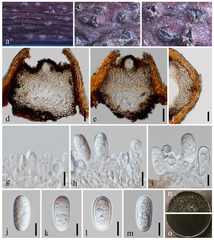
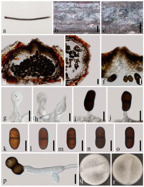
Diplodia mutila (GZAAS 23-0582). (a – c) Conidiomata on host substrate. (d, e) Vertical section of conidiomata. (f) Section of peridium. (g – i) Conidiogenous cells and developing conidia. (j – m) Conidia. (n, o) Colonies on PDA, above (n) and below (o). Scale bars: (d – f) = 20 μm, (g – m) = 10 μm.
Kredit dan penggunaan
© 2023 by the authors.
Ukuran dan format gambar disesuaikan tanpa pemotongan, retus, atau penambahan skala. Lisensi sumber dipertahankan.
Sumber profil
- [1] Botryosphaerialean fungi associated with woody oil plants cultivated in Sichuan Province, China — Diplodia mutila
- [2] Catalogue of Life Base2026-09-11 — Diplodia mutila
- [3] Dokumentasi visual — Diplodia mutila
- [4] Dokumentasi visual — Diplodia mutila
- [5] Dokumentasi visual — Diplodia mutila
Rujukan halaman ini
Fungi. Diplodia mutila. Rilis data fungi-2026-10-12-expansion-2. https://fungi-atlas.mgadib.chatgpt.site/jamur/diplodia-mutila/.
Status: impor sumber; pemeriksaan editorial per profil belum selesai. Untuk klaim biologis, gunakan rujukan pada bagian terkait dan periksa konteks sumber aslinya.