Ciri dan struktur
Notes: Our new isolates, MFLUCC 19–0335 (from MFLU 19–0544) and MFLUCC 17–2326 (from MFLU 17–1671), cluster with ex-type strain KUMCC 18–0090 of D. dehongensis, with strong support (100% MLBS/1.00 BIPP; Figure 1). Morphologically, the two new isolates fit well with the characters of D. dehongensis in having monoblastic or polyblastic conidiogenous cells and obpyriform to obclavate, straight or curved conidia with fewer than ten distosepta [ 45 ], except that the conidia of MFLU 19–0544 and MFLU 17–1671 show some percurrent proliferation, which is not observed in the holotype [ 45 ]. Additionally, the conidia of MFLU 19–0544 are pale-brown, while those of the holotype and MFLU 17–1671 are olivaceous. Comparisons of sequence data between the three strains showed no differences in LSU genes and three to six nucleotide differences in ITS genes. The holotype was collected from Yunan Provice, China [ 45 ], while our isolates were collected from Thailand, their locations constituting new geographic records. We provide new RPB2 sequence data for D. dehongensis in this study.
Habitat dan peran ekologis
Material examined: Thailand, Mukdahan Province, small river of Nong Bo Na Kae, found on dead, submerged, decaying wood of unidentified plants, 13 December 2018, Hao Yang, t34 (MFLU 19–0544), ex-type living culture MFLUCC 19–0335; Thailand, Long Khot subdistrict, found on submerged wood in a stream, 1 September 2017, Gennuo Wang, 4.7 (MFLU 17–1671), living culture MFLUCC 17–2326. Notes: Our new isolates, MFLUCC 19–0335 (from MFLU 19–0544) and MFLUCC 17–2326 (from MFLU 17–1671), cluster with ex-type strain KUMCC 18–0090 of D. dehongensis, with strong support (100% MLBS/1.00 BIPP; Figure 1). Morphologically, the two new isolates fit well with the characters of D. dehongensis in having monoblastic or polyblastic conidiogenous cells and obpyriform to obclavate, straight or curved conidia with fewer than ten distosepta [ 45 ], except that the conidia of MFLU 19–0544 and MFLU 17–1671 show some percurrent proliferation, which is not observed in the holotype [ 45 ]. Additionally, the conidia of MFLU 19–0544 are pale-brown, while those of the holotype and MFLU 17–1671 are olivaceous. Comparisons of sequence data between the three strains showed no differences in LSU genes and three to six nucleotide differences in ITS genes.
- Habitat atau konteks temuan
- kultur yang didokumentasikan
- Substrat
- Informasi belum tersedia pada sumber rilis ini.
- Peran atau konteks
- Belum dikategorikan pada rilis ini.
Reproduksi
Notes: Our new isolates, MFLUCC 19–0335 (from MFLU 19–0544) and MFLUCC 17–2326 (from MFLU 17–1671), cluster with ex-type strain KUMCC 18–0090 of D. dehongensis, with strong support (100% MLBS/1.00 BIPP; Figure 1). Morphologically, the two new isolates fit well with the characters of D. dehongensis in having monoblastic or polyblastic conidiogenous cells and obpyriform to obclavate, straight or curved conidia with fewer than ten distosepta [ 45 ], except that the conidia of MFLU 19–0544 and MFLU 17–1671 show some percurrent proliferation, which is not observed in the holotype [ 45 ]. Additionally, the conidia of MFLU 19–0544 are pale-brown, while those of the holotype and MFLU 17–1671 are olivaceous.
Catatan identitas dan cakupan
- Nama diterima mengikuti Catalogue of Life Base2026-09-11; uraian mempertahankan konsep dan nama sumber aslinya.
- Preserve original treatment localities, hosts and substrate scope. Materials examined describe documented specimens, not the full global range.
- Impor dan terjemahan otomatis belum diperiksa editorial per profil. Struktur reproduktif yang disebutkan tidak membuktikan seluruh siklus hidup diketahui.
- Pengukuran pada uraian mengikuti spesimen, kultur, dan metode sumber; angka tidak diperluas ke semua populasi.
Catatan distribusi dalam narasi mengikuti sumbernya. Dokumentasi spesimen atau foto mengikuti catatan sumber. Bukti wilayah Indonesia dan penilaian konservasi tidak disimpulkan dari gambar.
Melihat dengan konteks
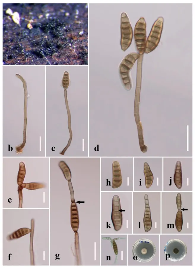
Distoseptispora dehongensis (from MFLU 19–0544). (a) Colonies on natural substrate. (b) Conidiophore. (c, d) Conidiophores with conidia. (e – g) Conidiogenous cells bearing conidia. Note the conidium with percurrent proliferation that is forming another conidium in g. (h – m) Conidia. Arrow in k points a gap in the middle of septum, which indicateds the distoseptate, note the percurrently proliferating conidium in m; (n) Germinated conidium. (o) Colony on PDA (from front). (p) Colony on PDA (from reverse). Scale bars: (b – c, n) 20 μm; (d, g) 30 μm; (e, f, h – m) 10 μm.
- Jenis
- Pelat ilmiah
- Kredit
- © 2022 by the authors.
- Lisensi
- CC BY 4.0
- Sumber media
- Buka entri sumber
Dokumentasi tambahan
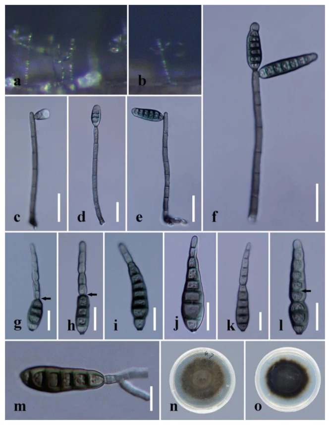
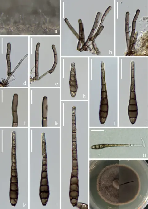
Distoseptispora dehongensis (from MFLU 17–1671). (a, b) Colonies on natural substrate. (c – f) Conidiophores with conidia. (g – l) Conidia. The arrows in g and h indicate conidial percurrent proliferating points. The arrow in l points to a gap in the middle of the septum, which indicates the distosepta. (m) Germinated conidium. (n) Colony on PDA (from front). (o) Colony on PDA (from reverse). Scale bars: (c – f) 20 μm; (g – l) 15 μm; (m) 10 μm.
Kredit dan penggunaan
© 2022 by the authors.
Ukuran dan format gambar disesuaikan tanpa pemotongan, retus, atau penambahan skala. Lisensi sumber dipertahankan.
Sumber profil
- [1] Polyphasic Identification of Distoseptispora with Six New Species from Fresh Water — Distoseptispora dehongensis
- [2] Catalogue of Life Base2026-09-11 — Distoseptispora dehongensis
- [3] Dokumentasi visual — Distoseptispora dehongensis
- [4] Dokumentasi visual — Distoseptispora dehongensis
Rujukan halaman ini
Fungi. Distoseptispora dehongensis. Rilis data fungi-2026-10-12-expansion-2. https://fungi-atlas.mgadib.chatgpt.site/jamur/distoseptispora-dehongensis/.
Status: impor sumber; pemeriksaan editorial per profil belum selesai. Untuk klaim biologis, gunakan rujukan pada bagian terkait dan periksa konteks sumber aslinya.